DR. DEPAEPE
1. WHAT?
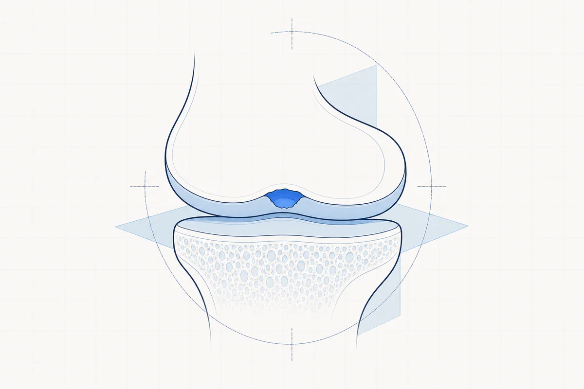
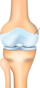
Articular cartilage is the mirror-smooth, white tissue that covers the ends of bones where they come together to form joints. This cartilage is a resilient tissue. After loading, the cartilage returns to its original shape, just like the action of a sponge. These properties make cartilage extremely suitable for absorbing pressure forces on the knee joint.
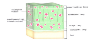
The joint surface is mainly made up of a special cartilage tissue called hyaline cartilage. Hyaline cartilage consists of approximately 90 percent water, which ensures its shape-restoring capacity. The hyaline cartilage is made up of four layers that gradually transition into bone. Hyaline cartilage contains less than 5 percent cartilage cells or chondrocytes. These create a network of collagen fibers. These fibers are woven together and always have a different direction in all four layers of the cartilage. This creates a strong structure. The top layer is somewhat deformable, so that no damage occurs when forces are exerted on it. The subsequent cartilage layers become firmer, so that they have a shock-absorbing effect. The deepest cartilage layer consists of the strongest collagen fibers. Underneath the 'real' cartilage there is a layer of calcium-rich cartilage. This forms the transition to the underlying bone.
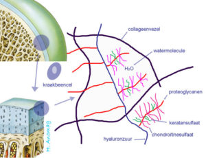
Proteoglycans are “trapped” in this cartilage meshwork. These large molecules are also made by the cartilage cells and want to bind water to them. This causes the proteoglycans to swell. This makes the cartilage stiffer, more elastic, smoother and stronger. The proteoglycans themselves are made up of glycosaminoglycans, including chondroitin sulphate, keratan sulphate and hyaluronic acid. This last substance also plays a role in the lubrication of the cartilage.
Cartilage has no blood flow, but it can be supplied with nutrients through load (pressure). Stress causes the cartilage to collapse, causing waste products to be pushed out of the cartilage. When the load is lifted, space is created to absorb water, oxygen and nutrients. It is therefore important to load and relieve cartilage. The nutrients are extracted from the synovial fluid. This fluid is continually replaced by movement of the joints, allowing the cartilage to absorb new nutrients. It is very important to keep moving, even if the cartilage is damaged.
2. FUNCTION
Articular cartilage ensures that both joint ends glide effortlessly over each other without friction. The articular cartilage functions as a shock absorber in the knee (together with the meniscuses).
3. CAUSE OF CARTILAGE INJURY
- Accident, sports accident: e.g. twisting movement of the knee. This is often accompanied by associated injuries to a meniscus or ligament. A sports accident is often responsible for an "isolated" cartilage injury: the adjacent cartilage still remains in good condition.
- Osteochondritis dissecans: This is a condition in which the underlying bone temporarily loses blood. The bone dies, causing the overlying cartilage to also die because it no longer has a stable surface. Osteochondritis dissecans is a disease that mainly affects children and young people.
- Overload: Frequent same movements with pressure on the same place in the knee gradually lead to wear and tear of the cartilage.
- Wear and tear: degeneration of the cartilage or the development of osteoarthritis. The relative content of collagen fibers increases and the amount of water decreases. This reduces the resilience of the cartilage. The result is that the cartilage layer starts to wear away and osteoarthritis develops.
- Rheumatic diseases.
However, cartilage is not provided with blood vessels, lymphatic vessels and nerve fibers. This means that it cannot be repaired if damaged. After all, blood flow is essential for repair of damaged tissue. A cartilage injury that is not treated has a greater chance of spreading.
4. COMPLAINTS OF A CARTILE INJURY
- Pain when straining the knee. Because cartilage does not have nerves and blood vessels that provide metabolism, shallow lesions usually do not cause pain. Complaints will often occur due to irritation of the synovium or mucous membrane of the knee. Once the cartilage lesions are deeper, the underlying bone will also become painful.
- Swelling.
- Cracking or grinding noise (crepitations).
- Blockage: if a loose piece of cartilage wanders around in the knee, this can cause blockages.
5. DIAGNOSIS AND INVESTIGATIONS
Clinical examination indicates whether there is fluid in the knee. With isolated cartilage injuries, the pain is often easy to localize.
Hyaline cartilage consists of approximately 90 percent water. As a result, cartilage cannot be visualized on an X-ray. Radiography does provide a very good estimate of the width of the joint space.
Arthro CT scan (CT scan with contrast fluid). Can clearly demonstrate the size of the injury.
MRI scan: can clearly demonstrate cartilage damage and is also important to demonstrate additional damage to other structures (e.g. meniscus).
Arthroscopy. This examination can best demonstrate the size of the lesions, the depth of the lesions, and the firmness of the edges of the lesions.
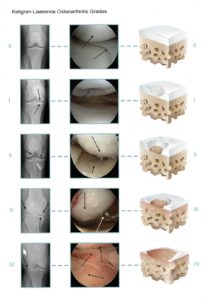
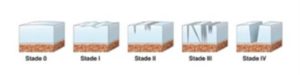
4 degrees of severity of cartilage injuries are described.
- grade 0: normal healthy cartilage
- grade 1: the cartilage shows a weak spot or blisters
- grade 2: small tears in the cartilage, but less than 50% of the thickness of the cartilage layer
- grade 3: lesions with deep pockets, involving more than 50% of the thickness of the cartilage layer
- grade 4: the deep tear in the cartilage exposes the underlying (subchondral) bone
6. TREATMENT
Traumatic, isolated cartilage injuries are quite treatable. Degenerative cartilage lesions are the most difficult to treat. Above the age of 50, there are few options for real surgical cartilage treatments. If cartilage injuries are not treated, the cartilage layer can gradually wear away, resulting in wear and tear and osteoarthritis.
6.1 Non-operative treatment
- Painkillers and anti-inflammatory medications.
- Weight reduction: Reducing the load on the knee joint will significantly reduce pain.
- Exercise therapy, physiotherapy: exercise sports that do not require body weight are excellent for the knee joint and provide nourishment to the cartilage (e.g. cycling).
- Cortisone preparation: to reduce pain and inflammation.
- Stem cells from abdominal fat.
6.2 Surgical treatment
Before starting treatment, your leg axis (O-leg or X-leg) will always be inspected. The leg axis can sometimes have a major effect on recovery after the treatment performed.
Arthroscopic debridement or “clean-up” operation:
During keyhole surgery, the loose cartilage pieces are removed using a shaver (mini cutter with vacuum cleaner). The edges of the cartilage damage are stabilized or made “smooth” again. Long-term results are difficult to predict.
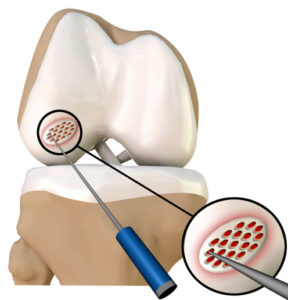
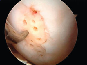
Microfracture or ice-pick surgery:
During keyhole surgery, an instrument (an awl or ice pick) is used to pierce small holes in the underlying bone. When the bone is punctured, the underlying bone marrow (with stem cells) comes through the holes in the injury. A blood clot containing stem cells forms in the injury that can fill the cartilage injury. These stem cells can then form a type of scar cartilage tissue (fibrocartilage). This scar tissue is always of inferior quality compared to the original hyaline cartilage.
This procedure has the best results in relatively young patients, with good underlying bone and isolated lesions up to 1.5 square centimeters. This technique still remains the gold standard for cartilage treatment.
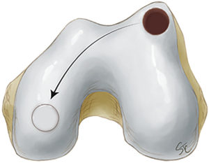
Mosaicplasty – OATS (Osteochondral Autograft Transfer System):
Plugjes kraakbeen met een deeltje bot eraan worden getransplanteerd van een gezond, niet-belast deel van de knie naar het letsel. De diameter en het aantal plugjes kan variëren naargelang de grootte van het letsel. Het letsel wordt dus opnieuw volledig opgevuld met lichaamseigen kraakbeen/bot. Gezien de hoeveelheid over te planten cilinders toch beperkt is, kunnen grote defecten vaak niet behandeld worden met deze vorm van kraakbeentransplantatie.
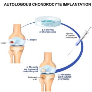
Cartilage cell transplantation (ACI – MACI):
In the first phase, pieces of cartilage are removed during keyhole surgery (biopsy). These are sent to the lab. The cells from these pieces are grown in the lab for about 10 weeks. In a second phase, these cultured cells are implanted through a small opening in the knee. This technique mainly aims at repairing the hyaline cartilage. This technique has now been almost completely abandoned due to disappointing clinical results and cost (not reimbursed).
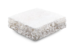
Cartilage scaffolds (Maioregen):
For injuries where both cartilage and bone are affected, an artificial cartilage membrane or scaffold can be implanted. This scaffold allows cartilage and bone to be repaired in the same procedure. Scaffolds look like a piece of foam or a plug and can be cut to size to fill the defect.
The top layer of a Maioregen consists of collagen and replaces the cartilage tissue. The bottom layer replaces the bone tissue. The material stimulates growth of new cartilage and bone tissue. Ultimately, the tissue will be largely absorbed by the body.
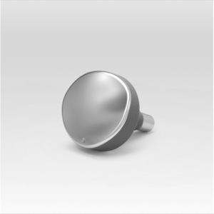
Metal implants (Episurf):
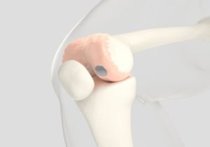
If larger cartilage lesions exist that cannot be resolved with the above biological solutions, a metal implant may offer a solution. Based on an MRI scan, the injury is reconstructed in 3 dimensions and a metal implant is provided to that size.
ANESTHESIA AND HOSPITAL STAY
The procedure is performed via day hospitalization or 1 night admission and can be performed via an epidural injection or short general anesthesia.
7. COMPLICATIONS
The chance of a complication during knee cartilage surgery is very small.
- Bleeding from the wounds or bleeding in the knee. We try to prevent this by applying lots of ice.
- The wounds can sometimes remain sensitive for a while after keyhole surgery. Sometimes there may also be a period of numbness around the scars.
- Persistent pain complaints. Realistic expectations are important! There is no biological cartilage treatment that can completely restore the injury to its original form.
- Infectie: zeer kleine kans omdat er uitvoerig wordt gespoeld tijdens kijkoperatie.
- A blood clot or phlebitis may form in your operated leg. We always give injections in the first week and we try to move as quickly as possible to reduce that chance to almost zero.
- Damage caused by keyhole surgery to structures around the knee, such as nerves or blood vessels, is very rare.
8. REHABILITATION
Because the treated area needs time to recover, the knee should only be reloaded very progressively. A specific physiotherapy schedule is given upon discharge from the hospital, depending on the technique used. During the first months of rehabilitation, a brace is also worn to relieve the treated area. There will always be a need to use crutches to limit support in the first 3 to 6 weeks.
9. FACTS
- Try the pain controlled with prescribed painkillers and ice applications.
- Love the wounds clean and dry. Showering is permitted with a shower plaster (opsite, tegaderm, etc.). The stitches can be removed after 14 days by the home nurse or your GP.
- Injections in the abdomen are recommended for the first 10 to 30 days (blood thinners to avoid phlebitis). This depends on the type of operation performed.
- Disability depends on the content of the job. The way you travel to work is also important. Discuss this with your doctor before the procedure.
- Driving. You can start driving when the knee feels reliable, strong and stable. It is also important that the knee bends at least 90°. For most people this is about 6 to 8 weeks after the procedure.
- Sports resumption. Your physiotherapist and surgeon will advise you when it is safe to resume your hobbies. This will depend on the type of sport and the level at which you practice it. Cycling is the ideal activity to make the knee flexible again and to train the thigh muscles without overloading the knee. After 4 to 6 weeks, non-load-bearing sports activities (e.g. cycling) can be progressively resumed if the pain and swelling have disappeared sufficiently. Intensive practice of running and jumping sports often takes 12 to 18 months.
Frequently asked questions
Orthopedisch chirurg – AZ Jan Palfijn Gent
Specialized in shoulder and knee surgery
The information on this page was prepared by Dr. Yves Depaepe and is intended as general medical information. Always consult your doctor for a personalized treatment plan.