DR. DEPAEPE
1. ANATOMY OF THE KNEE
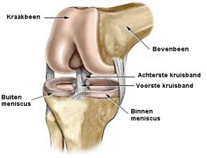
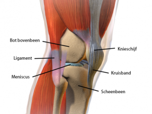
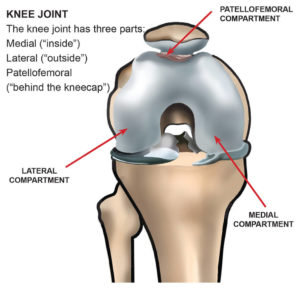
The knee is formed by 3 bones: the thigh bone (femur), the shin bone (tibia) and the kneecap (patella).
The ends of the femur, shinbone and the back of the kneecap are covered with cartilage.
This cartilage ensures smooth and painless rubbing of the joint surfaces opposite each other.
There are 2 shock absorbers between the femur and the shin bone, namely the inner meniscus (medial meniscus) and the outer meniscus (lateral meniscus).
Strong ligaments (medial collateral ligament, lateral collateral ligament, anterior cruciate ligament, and posterior cruciate ligament) connect the femur to the shin bone.
The knee is further controlled by the muscle groups of the femur and shin.
2. ARTHROSIS OF THE KNEE: cartilage wear and tear
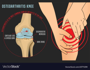
Osteoarthritis of the knee is a chronic wear and tear process in which the protective layer of cartilage at the bone ends gradually decreases in volume and quality.
In addition to the reduction in cartilage, the quality of the meniscus tissue also decreases with age. Cracks occur in the meniscuses as a result of wear and tear and they are less able to perform their shock-absorbing function.
As a result of the damage to the cartilage, the bony ends rub directly against each other, causing pain.
As a result of the wear and tear, a deformity of the knee will occur (O-leg or X-leg).
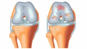
3. OTHER CAUSES of cartilage damage
- Osteoarthritis is the most common cause of cartilage disease (due to old age, due to accident, due to obesity).
- Rheumatoid arthritis is an inflammatory disease of the body that can also affect the knee and destroy its cartilage.
- avascular necrosis is a condition in which part of the bone dies. This leads to collapse of the bone under the cartilage, with progression to osteoarthritis.
4. COMPLAINTS of a “broken” knee
- increasing pain complaints when exercising, exercising, walking; cycling and swimming usually work better than walking.
- startstramheid, tijd nodig om ’s morgens terug soepel te worden.
- swelling of the knee.
- feeling of sagging of the knee.
- loss of mobility of the knee, reduced ability to straighten the knee.
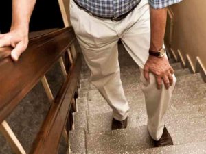
- general less active functioning: being unable to walk as far, being less able to climb stairs, finding it more difficult to make long car journeys, etc.
5. NON-OPERATIVE TREATMENTS
5.1 Medication
Analgesic and anti-inflammatory medications can be used to control symptoms.
5.2 Dietary supplements
Chondroitin and glucosamine are the most commonly used nutritional supplements. Turmeric is also frequently used.
The effect in large scientific studies has been rather limited to date.
5.3 Injections or infiltrations
Corticoids are most frequently used. These mainly provide an analgesic, anti-inflammatory and anti-swelling effect.
The number is usually limited to 3 per year.
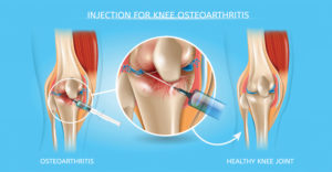
Hyaluronic acid (viscosupplementation) or “gel injections”
Hyaluronic acid is a gel-like substance that is normally present in healthy joints. It functions as a lubricant, shock absorber and thus protector of the cartilage layers.
In osteoarthritis, both the quality and quantity of this body's own hyaluronic acid are noticeably reduced. It becomes more difficult to nourish and protect the damaged cartilage
By artificially injecting, we try to control the symptoms of osteoarthritis. Repeatedly injecting hyaluronic acid could stimulate the production of the body's own hyaluronic acid. Usually 3 injections are necessary to achieve this result.
About 30% of treated patients become completely pain-free, while 20% will experience no effect.
The effect after the injections can last up to 18 months. If complaints recur, the injections can be repeated.
PRP or platelet-rich plasma
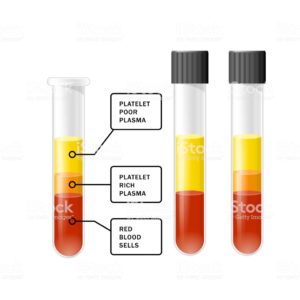
PRP uses the repair capabilities of our own platelets. The beneficial effects of these platelets were already known in the treatment of chronic tendon inflammation, but recently we have also noticed a beneficial effect on pain complaints, swelling and reduced functionality with injections in the knee for osteoarthritis.
After a blood collection, the plasma (with platelets) is separated from the other blood cells (white and red blood cells) via a centrifuge. The platelets are isolated and injected into the knee.
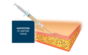
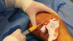
With stem cell therapy we attempt to control the pain caused by inflammation, stop the progression of cartilage breakdown and thus improve joint function.
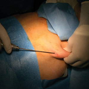
Classical liposuction of abdominal fat is performed using a standardized protocol to damage the stem cells as little as possible. The liposuction aspirate is washed several times in a centrifuge until a pure aspirate can finally be obtained that is injected intra-articularly (into the joint) into the knee.
5.4 Adjust lifestyle and activity
- keep moving frequently.
- cycle regularly.
- Avoid excess weight: when climbing stairs, up to 8x your body weight sometimes goes through the knee! A limited weight reduction can already lead to an improvement in complaints.
5.5 Physiotherapy
Learning specific exercises to strengthen muscle mass around the knee can lead to pain relief.
5.6 Knee bandage or brace
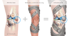
In specific circumstances this can lead to pain relief. Sometimes this is also prescribed to relieve the affected part of the knee for a period of time (unloader brace).
5.7 Arch supports
If an axis deviation of the leg occurs due to osteoarthritis, this can sometimes be corrected with an arch support. An arch support is in no way a treatment to prevent osteoarthritis.
6. TOTAL KNEE PROSTHESIS: a “new” or “false” knee
If the above treatments fail or if osteoarthritis is too advanced, we must proceed with complete or partial replacement of the knee joint.
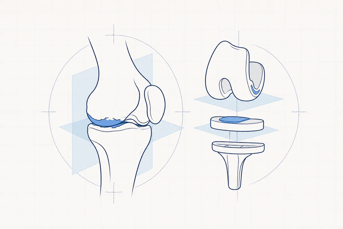
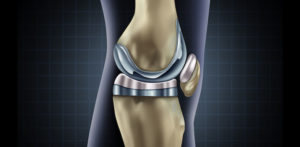
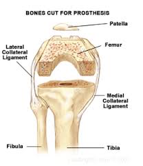
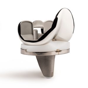
With a knee prosthesis, the entire knee is not removed! Only the damaged cartilage layers on the femur and shin are replaced by a special implant that perfectly mimics the natural shape of the original knee. The back of the kneecap is often also replaced with a new layer.
A plastic layer (wear-resistant polyethylene) is placed between the two metal layers as a new shock absorber. In the majority of cases, the implants are attached to the bone with a special bone cement (cemented prosthesis).
During the implantation of a prosthesis, the axis of the leg is also corrected to a normal position.
All this is done using a muscle-sparing technique with preservation of the lateral ligaments, which speeds up rehabilitation after surgery.
7. INNOVATION: Customized instrument (PSI: patient specific instrument)
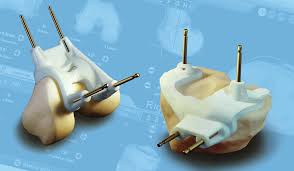
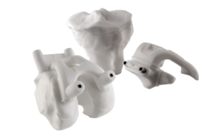
Nowadays it is possible to have a model of the knee made in advance using 3D scanners and 3D printing. Using this model, instruments are made to measure for your knee for single use. This partly facilitates the procedure and shortens the operating time, but is associated with an additional cost. This application can be very useful in special cases, but is by no means intended to be used for every operation.
8. INNOVATION: Robotic surgery
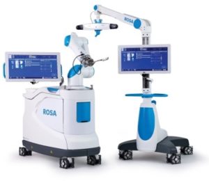
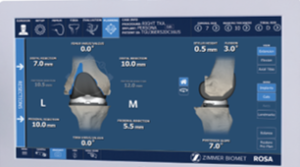
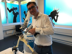
By switching on the robot, the position of the prosthesis can be individually adjusted to the specific anatomy of the patient. A 3D model of the knee is made in advance via special radiography and/or scan. Based on this 3D model, the robot can help us with very accurate cutting sections and the precise positioning of the components of the prosthesis. In this way we hope for even greater patient satisfaction and longer survival of the implant.
As a disadvantage we must mention a higher cost, but also a longer operating time, which can also entail complications (e.g. infection).
It may sound contradictory, but to date the benefit of using a robot to place a knee prosthesis has not yet been scientifically proven!
9. HALF KNEE PROSTHESIS or “half knee”
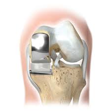
Only one part of the knee is damaged (inside or outside) and only this part receives a new surface. The parts of the knee that are not damaged do not receive a new layer.
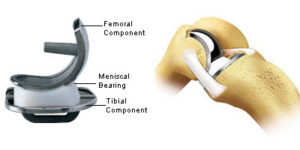
A half knee prosthesis consists of an implant on the upper leg and an implant on the lower leg. A hard layer of plastic (polyethylene) is applied between them to replace the shock absorber. With a 'half' prosthesis, the cartilage layer of the kneecap is not replaced. This prosthesis can be placed with or without bone cement, but usually the placement is without bone cement.
Het grote voordeel van dit type knieprothese is dat een groot deel van de eigen knie, inclusief de kruisbanden, kan behouden worden. Dit betekent voor de patiënten dan ook een snellere revalidatie en een knie die meer natuurlijk aanvoelt.
This procedure is of course only possible if only one compartment of the knee is affected and the other two compartments are intact.
10. PATELLOFEMORAL PROSTHESIS or kneecap prosthesis
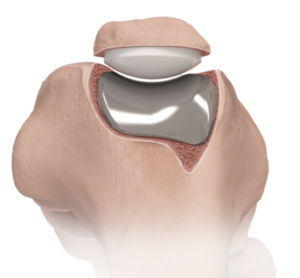
If the osteoarthritis occurs in the front compartment of the knee (the back of the kneecap and the groove in the thigh where the kneecap slides), such a prosthesis can be placed. It is essential to check in advance whether the complaints are limited to this compartment.
The trochlea (trough in the thigh) is replaced by a metal component and a plastic (polyethylene) component is placed on the kneecap. Both components are attached to the bone with bone cement.
11. ANESTHESIA or anesthesia for knee prosthesis
The procedure is performed under a short general anesthesia with associated local anesthesia via a locoregional block (Adductor Canal Block and LIA: local infiltration anesthesia). This provides better pain relief, less need for powerful painkillers (e.g. morphine derivatives) and less nausea. All this ensures that rehabilitation can start as quickly as possible (according to the rapid recovery protocol in our hospital).
In some medical circumstances, epidural anesthesia is chosen in combination with a light intoxication.
Your orthopedic surgeon will explain this to you before the operation. Of course, questions about the anesthesia can also be asked to the anesthetist.
12. COMPLICATIONS with knee prosthesis
Complications are inherent to any procedure. There is no such thing as a procedure without complications. As a rule, these rarely occur.
All precautions are taken as standard to keep the risk of complications very low.
Infections can occur superficially around the scar or deeper around the prosthesis. The infection can develop shortly after the procedure, but sometimes years later. Superficial infections can usually be treated with an antibiotic. Deep infections usually require major surgery with irrigation of the joint or sometimes removal of the prosthesis and long-term antibiotics afterwards.
If you have an infection anywhere in your body, it is wise to discuss this with your doctor quickly. It may be necessary to start antibiotics quickly to treat this infection and thus prevent the risk of infection via the bloodstream to the denture, e.g. tooth abscess.
Blood clot / Phlebitis. If these clots form in the lower leg, they can travel to the lungs and cause a life-threatening pulmonary embolism. As a preventative measure, injections or medication are always given during the first month to prevent this. Afterwards it is important to exercise enough, walking, cycling, etc.
Excessive scar formation: arthrofibrosis. Sommige mensen maken intern overmatig veel litteken aan. Dit geeft aanleiding tot meer stijfheid van de knie en moeizamere revalidatie. Het is daarom zeer belangrijk om zo snel mogelijk te starten met bewegen na de operatie en de oefentherapie nadien dagelijks vol te houden.
Allergy. This is almost non-existent and very rare. A special prosthesis with a coating can be placed in people with specific known metal allergies.
Nerve injury: superficial sensory disturbances around the scar are frequent; Serious nerve injuries such as foot drop when a nerve is damaged is extremely rare.
This list is not complete and can never be complete. There is always the possibility of developing a complication that is currently unknown or unproven in medical science.
13. PROSTHESIS SURVIVAL: How long will my prosthesis last?
Over the years, the plastic shock absorber (polyethylene) starts to wear slightly and the implants on the femur and tibia generally remain intact. With heavy activity and excess weight, the polyethylene wears out faster.
With current techniques and materials we expect a survival of approximately 20 years. This can change due to accidents, excessive strain, infections, diseases, etc.
When a knee prosthesis is worn out, in some cases a so-called revision operation will have to be performed. The old prosthesis is completely or partially removed and a new prosthesis inserted.
In a recent publication by the Lancet in 2019, approximately 82% of total knee arthroplasties and approximately 70% of unicondylar knee arthroplasties have a lifespan of 25 years.
14. SATISFACTION after placing the prosthesis
A large international study shows that more than 80% of patients are satisfied with the results.
The ultimate goal of our surgical technique is the “forgotten knee”.
Total knee arthroplasty is one of the most frequent and successful orthopedic operations. However, patient satisfaction in the short and long term after surgery can vary. Initially, the total knee prosthesis was developed for older patients with a limited life expectancy, but due to the continuous evolution in orthopedics with the development of newer and better materials, knee prostheses with the necessary reserve are also proposed to young active patients with severe osteoarthritis.
15. REHABILITATION
Following exercise therapy is essential for a good end result.
Make sure that sufficient pain medication is taken so that the exercise therapy can proceed comfortably.
The first phase of rehabilitation takes place in the hospital (about 4 days). After the operation, you may immediately put full weight on the new knee. In the initial phase this is sometimes difficult because of the wound pain. That is why you use crutches in this initial phase. According to a normal schedule, you use 2 crutches for 2 weeks, then switch to 1 crutch.
Afterwards, the rehabilitation takes place according to a given prescription. You can follow this rehabilitation at home with your own physiotherapist, in the hospital or in a rehabilitation center. 60 sessions of physiotherapy are prescribed.
De revalidatie kan in de beginfase enorm verschillen van patiënt tot patiënt. Dit heeft te maken met de ernst van de behandelde artrose, de grootte van de vooraf bestaande as afwijking en de toestand van de gewrichtsbanden. Hoed u om uw situatie te vergelijken met lotgenoten in de eerste weken na ingreep!
16. SPORT after a prosthesis?
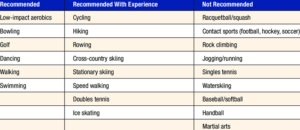
An active lifestyle such as walking, swimming or cycling is very important for maintaining good joint mobility. It is important to use your knee prosthesis carefully and sensibly. Avoid very strenuous exertion, avoid twisting movements in the knee joint, avoid frequent kneeling, such as e.g. while gardening. Always remember that this type of prosthesis has been developed for a normal active daily life without excessive strain.
Many people think that having a total knee prosthesis means the end of an active life and the end of any sporting activity. This is certainly not true! With a total knee prosthesis it is certainly possible to walk, swim, dance, intensively cycle, play golf, play tennis (doubles), ski, etc. But always use your common sense! Intensive and pivoting sports, such as basketball, long-distance running, water skiing, singles tennis, etc. are not recommended. These stressful sports will also reduce the survival time of your prosthesis.
17. COST
The reimbursement of the prostheses is closely monitored by the government. The highest quality is also guaranteed for every prosthesis! The majority of the prosthesis is covered by your health insurance company, but there is always a surcharge that is borne by the patient or the personal hospitalization insurance. It is best that you inform your health insurance company or insurer in advance. The charge for a knee prosthesis can vary depending on the type. The hospitalization insurance also intervenes to pay the surcharge that is not covered by the health insurance fund.
You can consult an average of a hospital invoice on the website of the Jan Palfijn hospital (cost estimate section).
https://www.janpalfijn.be/kostprijs-en-facturatie
18. FACTS after surgery
- How long in hospital? The recording takes 3 to 4 nights and sometimes varies individually. The wound must be sufficiently dry, the pain must be under control, the knee must be able to bend sufficiently and you must be able to use crutches and climb stairs easily.
- When can I shower? As soon as the wound is dry, a waterproof bandage can be applied, making showering possible!
- Swelling in the first weeks is completely normal. In some circumstances this can last up to several months. The swelling can be reduced by regularly applying ice and elevating your leg.
- Feeling warm in the knee. This can also last for several months and often depends on the activities you perform.
- Numb feeling around scar. Because the skin is incised at the front of the knee, only small nerve branches inevitably have to be cut. This zone of numbness corrects and diminishes over a period of 2 years.
- Click in my knee. This is the sound of the plastic shock absorber against the implant on the thigh. This occurs very frequently in the beginning. This becomes much less over the course of months.
- Driving: this depends on the speed of your individual rehabilitation and recovery of muscle strength. Driving can normally be resumed around 6 weeks after the procedure if you have sufficient control over your operated leg. Please consult the conditions of your car insurance in advance.
- Work disability. This is highly variable depending on the content of the job and varies from 6 weeks to 9 months.
- Kneel / sit on one knee is permitted but not comfortable, use a cushion if this does not provide sufficient comfort.
- Metal detectors at the airport. The metal detectors may indeed go off when you take the plane. However, the security services are used to this and quickly recognize that you have a prosthesis. You can always request an implant passport when you book a plane trip.
Frequently asked questions
Orthopedisch chirurg – AZ Jan Palfijn Gent
Specialized in shoulder and knee surgery
The information on this page was prepared by Dr. Yves Depaepe and is intended as general medical information. Always consult your doctor for a personalized treatment plan.