DR. DEPAEPE
1. ANATOMY
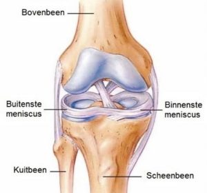
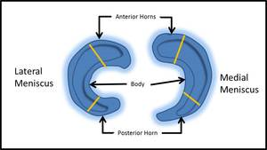
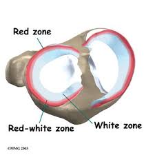
The meniscus is a semicircular, rubbery disc made of a special type of cartilage, which is located between the upper leg (femur) and lower leg (tibia). There is an inner C-shaped (medial or internal) and an outer 0-shaped (lateral or external) meniscus. Each meniscus consists of 3 parts: the posterior horn, the corpus (body) and the anterior horn. Three zones are also distinguished in the meniscus: an outer 1/3 zone (the red zone) with good blood circulation, a middle 1/3 zone (the red-white zone) with an average blood supply and an inner 1/3 zone (the white zone) without blood supply.
The inner meniscus is firmly connected to the capsule of the knee, the outer meniscus, on the other hand, is not tightly connected to the capsule and is therefore much more mobile.
2. FUNCTION
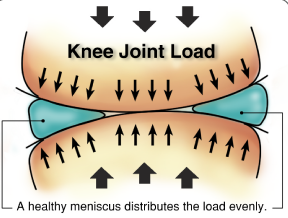
De meniscus heeft vooral een schok dempende functie. Door de schokken op te vangen wordt het bovenliggende en onderliggende kraakbeen beter beschermd. Daarnaast draagt een meniscus ook bij tot de stabiliteit van de knie (naast de gewrichtsbanden). Tevens heeft de meniscus een rol in de voeding van het kraakbeen dat de gewrichtsoppervlakken bedekt.
3. CAUSE OF MENISCUS TEAR
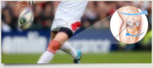
An incorrect movement, such as a twisting movement from a squatting position or a sudden twisting movement during sports, can cause a meniscus tear (traumatic meniscus tear). The meniscus can also tear in the event of a very minor trauma, if it has already been significantly weakened due to a previous wear process (unraveling) (degenerative meniscus tear or wear tear).
Meniscus tears occur in every age category. In the younger group, it is more likely to involve isolated traumatic meniscal injuries. In the older group, this mainly concerns degenerative cracks and there will be frequent accompanying cartilage lesions (wear and tear). The latter determine whether or not the knee will become pain-free after treatment.
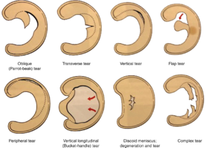
The type of accident and the quality of the meniscal tissue determine which type of tear injury occurs.
4. COMPLAINTS OF A MENISCUS TEAR
Not every meniscus tear causes complaints! The load depends on the type of crack and the location of the crack. The body weight, the axis of the leg and the degree of physical activity also determine the complaints.
- Pain: is mainly present during deep flexion and full extension of the knee. This pain decreases little by little after the accident.
- Stiffness: is usually most present in the first period after the accident.
- Swelling: is the result of fluid accumulation or bleeding within the knee.
- Blocking: If part of the meniscus becomes wedged between the upper leg and lower leg, the knee can no longer be fully bent or straightened.
- Sagging feeling: feeling of not standing firmly on your leg and bending at the knee.
- “Click”: sometimes there is a palpable or audible click in the knee joint.
5. RESEARCH
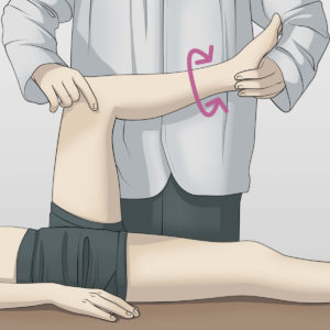
A good one clinical research from the doctor may suggest the diagnosis.
Radiography: of the knee gives us an idea of the amount of wear and tear on the joint.
MRI scan: is often the appropriate examination to visualize a meniscus tear.
CT scan with contrast fluid (arthro-CT) is sometimes used if significant additional cartilage damage is already suspected.
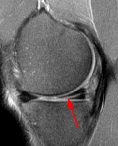
6. TREATMENT
The treatment depends on the type of tear, the size of the tear, the location of the tear, the quality of the meniscus tissue and the age. Not every meniscus tear is treated surgically.
In young people, accidental tears are treated with keyhole surgery. In the group of “degenerative tears” in patients over 50 years of age, an attempt is made to distinguish between “stable” and “non-stable” meniscal tears. The first treatment for stable degenerative tears will be non-surgical. If complaints still persist after months, a decision may still be made to perform keyhole surgery.
NON-OPERATIVE TREATMENT
- Rest, ice applications.
- Anti-inflammatory medication to reduce pain and inflammation.
- Injection with a cortisone preparation to reduce pain and inflammation, with hyaluronic acid to protect cartilage or sometimes with PRP.
- Muscle strengthening exercises and physiotherapy.
OPERATIVE TREATMENT: EXPOSURE OPERATION OR ARTHROSCOPY OF THE KNEE
A camera and several specific instruments are inserted into the knee through two small openings of a few millimeters. During arthroscopy, the joint is filled with fluid, so that there is more space in the knee and the joint can be continuously flushed. The purpose of keyhole surgery is not just to look, but also to solve the problem!
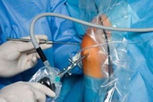
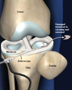
Partial or partial menisectomy
If the meniscus has suffered irreparable damage, the tear is removed during keyhole surgery with special instruments. This mainly concerns tears in the inner part of the meniscus where there is no or insufficient blood supply (white zone, see section 1). Naturally, only the damaged part is removed, the remaining part of the meniscus is preserved. In this way, a large part of the shock absorber is retained and the risk of osteoarthritis is kept to a minimum. This is the most commonly performed arthroscopic procedure in the world with favorable results.
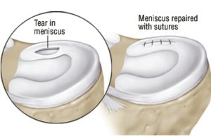
Meniscus suture
Recent tears in the red, well-vascularized area (see section 1) are candidates for suturing. The decision to suture is often made during keyhole surgery. The torn fragments are stitched back together using special anchors and wire.
The major advantage of a suture is that the meniscus can retain its full function. The disadvantage is that the meniscus needs time to heal, which means there are certain restrictions during the first 3 months after surgery. This is to give the meniscus the opportunity to grow back into place. This bonding should be seen as a long-term investment.
Jammer genoeg genezen meniscushechtingen slechts in maximaal 8 op de 10 gevallen, waardoor dus soms later nog een heringreep nodig is.
Artificial meniscus or meniscus scaffold
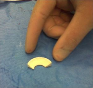
If a large part of the meniscus is lost, consideration is sometimes given to replacing this. One of the options is to implant a type of artificial meniscus into which the body's own cells can grow and develop into meniscus-like tissue. This treatment is currently mainly used in a study context and is certainly not a frequently used technique.
Meniscal transplant
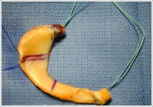
In young people where a severely damaged meniscus had to be almost completely removed and where the cartilage is still completely intact, a meniscus transplant is sometimes considered. The entire meniscus is replaced by a donor meniscus via keyhole surgery. There are often long waiting lists to find a suitable meniscus donor.
ANESTHESIA AND HOSPITAL STAY
The procedure is performed as a day hospital and can be done via an epidural or short general anesthesia.
7. COMPLICATIONS
- Bleeding from the wounds or bleeding in the knee. We try to prevent this by applying lots of ice.
- The wounds (no matter how small) can sometimes remain sensitive for a while after keyhole surgery.
- Persistent pain complaints. Sometimes there are associated cartilage lesions associated with degenerative meniscus tears that cannot be resolved with keyhole surgery. This will be discussed with your doctor upon consultation.
- Infection: very small chance because extensive rinsing is done during keyhole surgery.
- A blood clot or phlebitis may form in your operated leg. We always give injections in the first week and we try to move as quickly as possible to reduce that chance to almost zero.
- Damage caused by keyhole surgery to structures around the knee, such as nerves or blood vessels, is very rare.
8. REHABILITATION
Partial or partial menisectomy
It is best to use crutches for 5 days to give the knee a rest and avoid swelling. Afterwards, support is fully permitted.
During your stay at the day hospital, you will be taught exercises that you can perform every day. If necessary, you will receive a prescription for physiotherapy.
As soon as cycling is possible, you can cycle unloaded, for example on an exercise bike.
Meniscus suture
During the first 3 weeks you will have to walk between crutches without fully supporting your leg. Only the toes should touch the ground. The physiotherapist will teach you how to use the crutches.
You will receive a prescription for physiotherapy and these guidelines must be followed.
Deep folding beyond 90° is not recommended for the first 6 weeks to limit tension on the suture. A knee brace is usually used to prevent it from bending over 90°.
9. FACTS
- Try to control the pain with prescribed painkillers and ice applications.
- Keep the wounds clean and dry. Showering is permitted with a shower plaster (opsite, tegaderm, etc.). The stitches can be removed after 14 days by the home nurse or your GP.
- Injections in the abdomen are recommended for the first 10 days (blood thinners to avoid phlebitis). You will receive a prescription for home care, the nurse at the day hospital can also teach you how to do it. If you are already taking blood thinners before, this may differ from the normal schedule.
- Incapacity for work is between 3 days and 6 weeks and depends on the content of the job. The way you travel to work is also important. Discuss this with your doctor before the procedure.
- You can start driving when the knee feels reliable, strong and stable. It is also important that the knee bends at least 90°. A normal gait and walking down stairs without using the handrail provide a good indication of muscle control around the knee. For most people this is about 1 to 3 weeks after the procedure.
- Sports resumption. Your physiotherapist and surgeon will advise you when it is safe to resume your hobbies. This will depend on the type of sport and the level at which you practice it. Cycling is the ideal activity to make the knee flexible again and to train the thigh muscles without overloading the knee. Sports activities can be resumed gradually after 4 to 6 weeks if the pain and swelling have disappeared sufficiently.
- These facts apply to a partial menisectomy. The guidelines after meniscus suturing or other meniscus procedures may differ significantly and will be discussed with you by the doctor.
Frequently asked questions

Orthopedisch chirurg – AZ Jan Palfijn Gent
Specialized in shoulder and knee surgery
The information on this page was prepared by Dr. Yves Depaepe and is intended as general medical information. Always consult your doctor for a personalized treatment plan.