DR. DEPAEPE
1. Anatomy
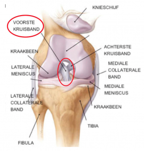
The anterior cruciate ligament is one of the four major ligaments of the knee.
The ligaments are four cord-like structures that connect the upper leg (femur) to the lower leg (tibia).
They are the most important stabilizers of the knee and ensure that the femur does not slip off the tibia.
The anterior and posterior cruciate ligaments are located centrally within the knee. The inner (medial collateral ligament) and outer ligament (lateral collateral ligament) lie on the inside and outside of the knee, respectively.
2. Function of the anterior cruciate ligament
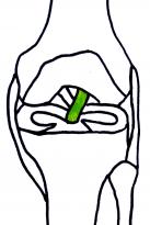
The anterior cruciate ligament provides fore-aft stability of the knee.
The anterior cruciate ligament prevents the lower leg from moving abnormally forward compared to the upper leg.
The anterior cruciate ligament also plays an important role in rotational control of the knee and thus protects the meniscus.
3. Cause of cracks
70% of anterior cruciate ligament tears are the result of non-contact mechanisms.
Usually there is a turning maneuver with a stretched knee and blocked foot.
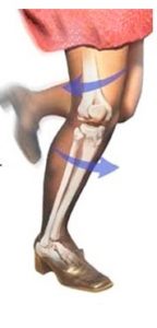
In football there is the typical “cutting” maneuver. The player makes a pivoting motion over the supporting leg to change direction.
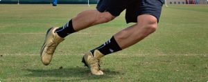
Poor landing after a jump, e.g. basketball, volleyball, are also frequent causes of a tear.
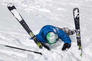
Ook tijdens het skiën als de binding van de ski niet tijdig loskomt ontstaat een draaibeweging in de knie.
30% of torn cruciate ligaments are the result of direct contact, such as a tackle in football.
Several studies have shown that female athletes are more susceptible to anterior cruciate ligament injuries (including differences in muscle strength and control and the effect of estrogen on the strength of the cruciate ligament).
4. Symptoms
The patient very frequently mentions that he has felt or heard a “crack”.
Gradually more swelling of the knee occurs (due to bleeding within the knee) and stiffness develops that makes walking uncomfortable.
There are often additional ligament or meniscus injuries that influence the pain and complaints.
In the second phase, the knee becomes flexible again and an "uncertain" or sagging feeling arises, for example during exercise or descending stairs.
Sometimes the symptoms are mild, which can make diagnosis difficult.
5. Diagnosis
1. Based on the patient's story, the doctor tries to reconstruct the accident mechanism.
2. Clinical examination by the doctor.
The anteroposterior stability and rotational stability of the knee will be tested through a number of specialized tests.
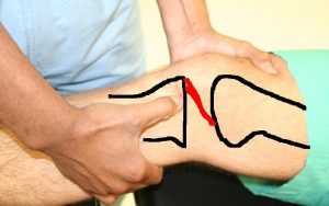
These tests are not always easy to perform immediately after trauma because of the pain.
3. Imaging
An emergency radiography is often performed to rule out bony injuries.
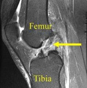
In most cases, an MRI scan is indicated to confirm the tear and to detect additional injuries to cartilage, meniscus and ligaments.
6. Healing? Recovery? Why treat?
Unlike other ligaments, a torn anterior cruciate ligament will not recover spontaneously.
As a result, there is a loss of stability in the knee joint with the risk of sagging during sports or stressful activities.
In the long term, there is a greater risk of cartilage damage, meniscus tears and osteoarthritis of the knee.
With untreated anterior cruciate ligament injuries, 90% of patients will experience meniscal damage within 10 years.
7. Treatment in acute phase
Rest is recommended in the first 2 to 3 weeks.
Ice applications are necessary to control swelling.
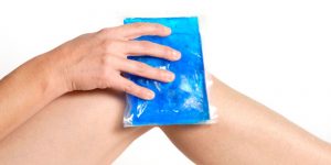
Anti-inflammatories are often prescribed in the first 2 weeks.
As soon as the knee is sufficiently deswollen and the pain permits, physiotherapy is started. The aim of physiotherapy in the first phase is to make the knee flexible again (full flexion/extension). Muscle strengthening exercises follow in the second phase.
8. Non-operative treatment
Does every anterior cruciate ligament tear need to be treated surgically?
In the minority of cases, an anterior cruciate ligament injury will be treated non-operatively.
The emphasis of the treatment is then to follow a muscle-strengthening exercise program.
This non-operative treatment is indicated for patients who lead an inactive life (e.g. sedentary work, no sports, etc.) and who do not experience a lasting feeling of sagging or instability.
If there are additional cartilage or meniscus injuries, surgery is usually also indicated.
9. The procedure
The aim of the procedure is to obtain a stable knee.
In the majority of cases, reconstruction is performed with your own tendons, donor tendons or artificial tendons.
Seal of an anterior cruciate ligament tear is only possible in a very small number of tears.
The procedure with reconstruction is best performed 4 to 6 weeks after the accident.
It is especially important that the knee is sufficiently deswollen and mobility is restored. This will undoubtedly speed up the rehabilitation afterwards.
The procedure is performed under a short general anesthesia and additional local anesthesia (femoral block, Hunter canal block).
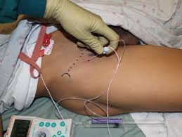
The procedure is always performed via keyhole surgery.
10. Anterior cruciate ligament reconstruction
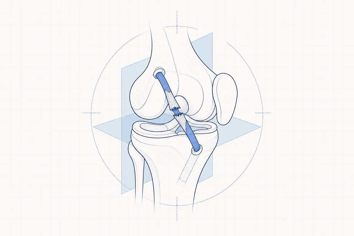
The torn cruciate ligament is replaced by a new sturdy structure, usually a tendon.
This tendon can come from your own body: hamstring tendons (on the inside of the knee), part of the patellar tendon (tendon under the kneecap) or part of the quadriceps tendon (tendon above the kneecap).
In specific circumstances, the tendon from the donor bank/donor tendon or a synthetic cord (e.g. LARS ligament) can be used.
The hamstrings are used most frequently.
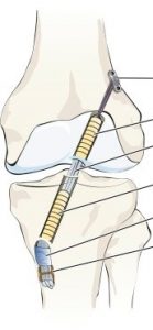
During keyhole surgery, the old remains of the torn cruciate ligament are removed. Two tunnels are drilled, one in the tibia and one in the femur. Both tunnels are positioned at the location of the original cruciate ligament. In exceptional cases, multiple tunnels can be used (double bundle technique).
The own tendon is prepared with a special fixation thread.
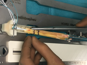
The whole is brought into the knee via the drill tunnels.
The new tendon is suspended in the tunnel of the femur. Classically, an endobutton system is used.
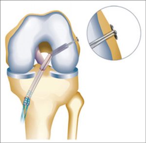
Afterwards, the fixation takes place with the correct tension on the shin bone.
Usually a resolvable screw is used (these do not have to be removed later, but disappear on their own after a while).
Bijkomend kan op het scheenbeen een schroef, kram, anker geplaatst worden ter versterking van de fixatie.
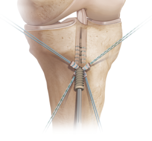
It is important that the new cruciate ligament is in the same position as the old original cruciate ligament.
The fixation system is very important because it is responsible for the stability until the own tendon has grown into the bony tunnels. The transplanted tendon is converted into living ligamentous tissue. This process usually takes at least 6 months.
In some cases there is a need for additional extra stabilization for rotational stability. This additional procedure is mainly used in risk groups (pronounced instability, revisions, highly competitive pivot athletes, hyperlaxity, etc.)
In this case, a monoloop procedure (lateral extra-articular tenodesis) or anterolateral ligament reconstruction is often additionally performed.
This will be discussed with the treating physician before the procedure.
11. Hospital stay
After the procedure you will stay in hospital for 1 night.
The physiotherapist in the hospital will teach you how to use the crutches and will teach you some exercises to do at home.
12. Homecoming
There is often still a lot of swelling of the knee in the first phase. This is completely normal. Frequent ice applications and frequent elevation of the leg can reduce this.
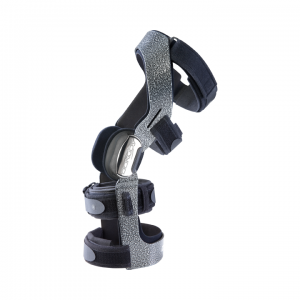
A brace for the first 6 weeks is necessary.
Make an appointment for the physiotherapist for the day following your return home.
The wounds from keyhole surgery must be cared for cleanly and kept dry. A waterproof bandage can be applied before showering (e.g. Opsite)
An injection is required for 20 days to prevent phlebitis.
If you prefer not to administer this injection yourself, you can contact a home nurse.
If you are already taking blood thinners before, this may differ from the normal schedule.
13. Rehabilitation
This phase of the treatment is just as important as the procedure itself.
The rehabilitation takes place under the supervision of a physiotherapist in accordance with a given prescription.
The procedure is protected during the first 6 weeks of rehabilitation by wearing a brace.
The new tendon is also protected by using crutches in the first 4 weeks. This will be reduced with the physiotherapist according to the schedule and according to the progress of the rehabilitation. (mobility, muscle strength,…).
14. What/when after surgery?
The sport-specific rehabilitation is discussed with the treating physician and, if necessary, also with the coach or club manager.
Frequently asked questions
Orthopedisch chirurg – AZ Jan Palfijn Gent
Specialized in shoulder and knee surgery
The information on this page was prepared by Dr. Yves Depaepe and is intended as general medical information. Always consult your doctor for a personalized treatment plan.