DR. DEPAEPE
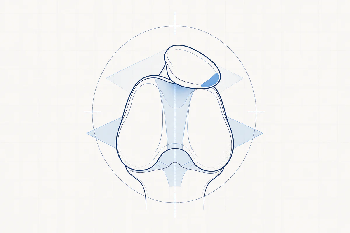
The patellofemoral joint is the joint between the kneecap and femur.
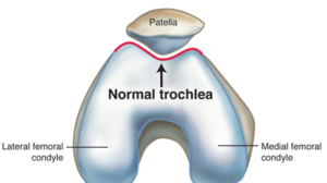
The kneecap is connected to a strong tendon in the lower leg (patellar tendon). At the top, the kneecap is attached to the four-headed thigh muscle (quadriceps). This muscle pulls the kneecap over the thigh, causing the knee to straighten. The V-shaped kneecap runs in a V-shaped groove/groove of the upper leg (trochlea of the femur) and forms the patello-femoral joint.
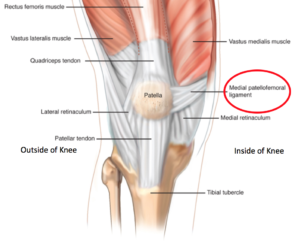
An MPFL or Medial Patello Femoral Ligament is a ligament on the inside of the knee and runs from the kneecap to the inside of the thigh. This ligament ensures that the kneecap stays in place in the middle of the knee in the intended groove and does not 'jump' or luxate out of its groove.
The patellofemoral joint is a complex joint where various problems can cause pain.
- stability problems: the kneecap can “jump” out of the gutter.
- patellofemoral pain syndrome
- cartilage injuries and osteoarthritis pain of the kneecap
PATELLA INSTABILITY or PATELLA LUXATION
1. WHAT
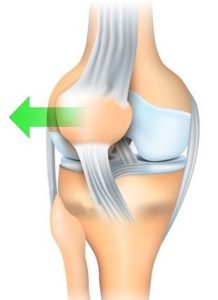
The normal kneecap is located centrally in relation to the knee and moves up and down in a kind of groove when making flexion and extension movements.
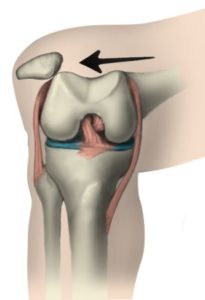
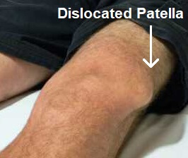
If the kneecap does not "track" properly in that groove in the thighbone, it can tend to derail from the groove, or even actually pop out of the socket (patellar luxation).
2. CAUSE
The cause is usually traumatic, most typically in a sports accident.
However, there are often underlying, predisposing factors that make it easier for the kneecap to “pop out of the gutter”.
- Abnormal shape of the gutter (dysplasia)
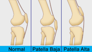
- Kneecap implanted too high (patella alta)
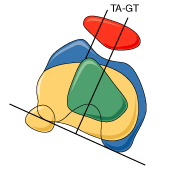
- Abnormal placement of the patellar tendon on the lower leg (increased TAGT value)
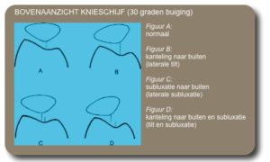
- Tilting of the kneecap to the outside (increased patella tilt)
- Hyperlaxity, overly mobile joints
During a patellar luxation, the Medial Patellofemoral Ligament (MPFL) tears, increasing the risk of a subsequent patellar luxation.
If multiple risk factors are present, the risk of dislocation (luxation) of the kneecap increases.
3. COMPLAINTS
- Pronounced pain during acute dislocation. This pain will only decrease when the kneecap is back in place (reduced). Sometimes this reduction must be done by the (emergency) doctor.
- Significant swelling after the dislocation because there is blood in the knee due to a tear in the MPFL ligament (hemarthrosis).
- Gevoel dat knieschijf telkens weer “uit kom” zou schieten
4. RESEARCH
- Radiography: shows the shape of the “gutter” and any associated fractures.
- NMR shows bone edema (fluid in the bone) at the lateral femoral condyle and/or medial facet of the kneecap. The NMR can show loose cartilage fragments and can confirm that the MPFL ligament is torn.
- CT Scan: to visualize the predisposing factors (dysplasia, patella alta, increased TAGT, patella tilt), a CT scan is the most accurate.
5. TREATMENT
NOT OPERATIVE
After an initial dislocation, a plaster cast and/or brace is worn for about 3 weeks. This is followed by intensive exercise therapy with the physiotherapist to strengthen the muscles around the kneecap.
OPERATIVE
In case of multiple dislocations, repeated dislocations or clearly predisposing factors, surgery should be considered. Every time a dislocation occurs, additional damage is caused to the knee!
MPFL RECONSTRUCTION
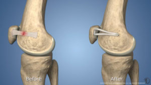
The MPFL ligament always tears when the kneecap is dislocated. Repairing this ligament is therefore the most essential surgical treatment.
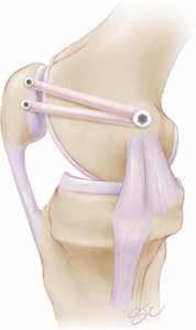
The torn MPFL ligament is reconstructed with its own tendon (hamstring tendon). This is very similar to the tendons used in anterior cruciate ligament repair. The new tendon is anchored on the inside of the kneecap with 2 screws and on the inside of the thigh with 1 screw. In this way, a new strong ligament is created with the correct tension that prevents the kneecap from luxating again.
In some patients, cartilage damage may occur during a dislocation. In most cases, the cartilage damage will heal spontaneously by restoring stability through MPFL reconstruction.
TUBEROSITY OSTEOTOMY AND TRANSFER
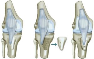
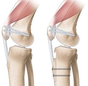
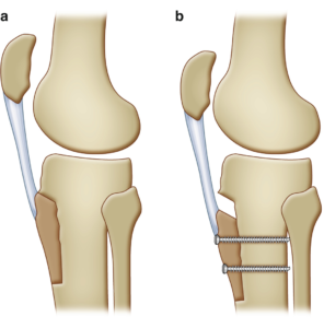
If the kneecap is too high (patella alta) or if the implantation of the kneecap tendon on the lower leg is too far outward (too high TAGT), an MPFL reconstruction alone is often insufficient and an additional procedure must be performed.
At the point where the patellar tendon attaches to the lower leg (tuberosity), the tendon is released with a bone block. This bone block with the patellar tendon attached to it is moved and reattached in the optimal location using 2 screws.
ANESTHESIA AND HOSPITAL STAY
The procedure is performed via an overnight stay and can be performed via an epidural or short general anesthesia. An additional injection in the groin is often given as temporary local anesthesia to reduce pain after the procedure.
6. COMPLICATIONS
Complications can arise with any procedure. The chance of a complication with this procedure is rather small.
- Bleeding from the wounds or bleeding in the knee.
- The wounds sometimes remain sensitive for a long time or the skin around the wounds feels temporarily numb.
- A thrombosis or pulmonary embolism may occur. As a preventive measure, injections (blood thinners) are always prescribed so that this risk is extremely small.
- Infection around knee joint. Good wound care is essential. An antibiotic is administered during the procedure to keep this chance very small.
- Damage caused by the operation to structures around the knee, such as nerves or blood vessels, is very rare.
- New tear of the repaired MPFL ligament. This is not really a complication, the kneecap can of course dislocate in the event of a new accident.
- Failure of the bone to fuse with a tuberosity osteotomy. Smoking cessation is absolutely recommended to achieve faster healing.
- Irritation of the subcutaneous screw heads during tuberosity osteotomy. If these are bothersome (e.g. when kneeling), they can be removed via a minor procedure once everything has healed.
7. REHABILITATION
MPFL
Depending on the pain, the knee may be immediately bent and straightened.
Support is immediately permitted between two crutches. After 3 weeks, the crutches can be gradually reduced.
The exercise therapy is done via a prescribed physiotherapy schedule with your own physiotherapist.
Tuberosity osteotomy
The mobility of the knee is limited by a knee brace with a hinge. According to the provided schedule, your physiotherapist will adjust the hinge of the brace so that you can progressively move more.
Support is severely limited in the first 3 weeks (only with toes on the floor) and between crutches. After 3 weeks, support will be gradually practiced. This step pattern is taught to you by the physiotherapist.
The exercise therapy is further carried out via a prescribed physiotherapy schedule with your own physiotherapist.
8. FACTS
- Swelling in the first few weeks is completely normal. In some circumstances this can last up to several months. The swelling can be reduced by regularly applying ice and elevating your leg.
- When can I shower? As soon as the wound is dry, a waterproof bandage can be applied, making showering possible!
- Sutures: the sutures can be removed 2 weeks after the procedure.
- Numb feeling around scar. Because the skin around the knee is incised, some skin branches inevitably have to be cut. This zone of numbness corrects and diminishes over a period of 2 years.
- Driving: this depends on the speed of your individual rehabilitation and recovery of muscle strength. It is also important that the knee bends at least 90°. Driving can normally be resumed around 6 weeks after the procedure if you have sufficient control over your operated leg. Please consult the conditions of your car insurance in advance.
- Incapacity for work: this is very dependent on the job content and varies from 3 weeks to 6 months.
- Return to sports: your physiotherapist and surgeon will advise you when it is safe to resume your hobbies. This will depend on the type of sport and the level at which you practice it. Cycling is the ideal activity to make the knee flexible again and to train the thigh muscles without overloading the knee. After 4 to 6 weeks, non-load-bearing sports activities (e.g. cycling) can be gradually resumed if the pain and swelling have disappeared sufficiently. More intensive practice of running and jumping sports often takes 4 (MPFL) to 8 (tuberosity osteotomy) months.
PATELLOFEMORAL PAIN SYNDROME
1. WHAT
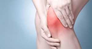
This concerns pain around the kneecap. Patellofemoral pain syndrome occurs most often in girls and young women (12-25 years).
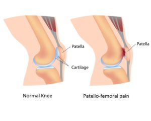
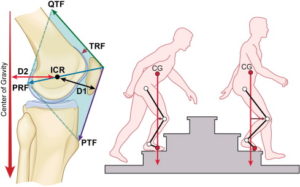
The pain usually manifests itself when there is pressure on the kneecap. During activities such as climbing stairs, descending and jumping, the kneecap has to handle up to eight times the body weight. Static activities such as standing for a long time, sitting for a long time with deeply bent knees and squatting also build up enormous pressure in the knee joint.
2. CAUSES
There are many different factors that can cause patellofemoral pain syndrome:
- X-legs
- rotational abnormalities in the lower limbs
- shortened muscles and/or tendons
- disturbed functioning or development of the muscles around the kneecap
- abnormal shape of the gutter in the thigh (trochlea)
- abnormal shape of the kneecap
- abnormal placement of the patellar tendon on the lower leg
- hyperlax joints
- overload of the knee or post-traumatic (e.g. impact of the knee)
- overweight and/or knee-taxing activities (e.g. squatting, climbing stairs, intensive sports)
- cartilage damage to the kneecap or the gutter in the thigh
Als één of meerdere van bovenstaande factoren aanwezig zijn, wordt de behandeling van de klachten daaraan aangepast. In de meeste gevallen van het patellofemoraal pijnsyndroom wordt er echter geen duidelijke oorzaak gevonden en is het meer irritatie van de structuren rondom de knieschijf die leiden tot de klachten.
3. COMPLAINTS
The complaints of patellofemoral pain syndrome are very diverse. It is often a combination of two or more of the following complaints:
- a constant feeling of pressure around the kneecap (“feeling the knee”)
- stiffness of the knee with sporadically limited swelling
- pain when going up or down stairs
- pain when kneeling, squatting, standing/sitting for a long time (e.g. receptions, cinema)
- pain when cycling uphill or walking downhill
- Cracking feeling or stuttering of the knee when bending/extending
4. EXAMINATIONS AND DIAGNOSIS
The diagnosis of patellofemoral pain syndrome is made based on the patient's pattern of complaints and clinical examination.
The clinical examination may show that additional research is indicated to identify possible causal factors.
An X-ray of the knee provides a clear picture of the anatomy of the kneecap and its relationship to the groove in which it tracks.
If there are suspicions of cartilage damage or if specific measurements need to be taken, an MRI or CT scan may also be indicated.
5. TREATMENT
The complaints are very annoying, but they often disappear spontaneously in adulthood. Contrary to what is sometimes claimed, patellofemoral pain syndrome is not a precursor of osteoarthritis!
Most complaints must be treated conservatively (i.e. not surgically) by one or more of the following treatments:
- Physiotherapy: Exercise therapy, in the form of physiotherapy in combination with home exercises, is the foundation of the treatment. The patient receives a specific physiotherapy schedule to correctly stretch and train the muscles of the knee with extra attention to certain muscle groups, which are very important for the correct tracking of the kneecap in the groove.
- Relative peace: It is often sufficient to take a period of (relative) rest or temporarily reduce sporting activities to a lower level or lower intensity. Sometimes it is necessary to temporarily stop the sport completely and first train certain muscles with the physiotherapist. The sport can then be resumed under supervision.
- Small tools can sometimes make a big difference, for example you can raise the saddle of the bicycle slightly to reduce kneecap pain. Cycling with light gear works better than with heavy gear.
- It may also be advisable to temporarily avoid movements that provoke pain (kneeling, standing for a long time, climbing stairs, sitting in a folded position for a long time, etc.) until the knee is sufficiently trained through exercise therapy to tolerate these movements again.
- Anti-inflammatories and ice: Frequently applying ice to the kneecap in combination with a short course of an anti-inflammatory (NSAID) has an analgesic and anti-inflammatory effect.
- Weight loss: Bij patiënten met overgewicht kan een gering gewichtsverlies (bv. 5 kg) al voldoende zijn om de klachten te verbeteren. Zoals hierboven al vermeld, moet de knieschijf bij activiteiten zoals trappen lopen tot acht keer het lichaamsgewicht verwerken. 5 kg minder lichaamsgewicht wordt bijgevolg door de knie aangevoeld als 40 kg minder druk.
- Cartilage supplements: In certain cases, nutritional supplements may be prescribed. This is not a medication, not harmful and can be taken for a long time without side effects. A treatment must be followed for several months to evaluate its effect. Cartilage supplements obviously cannot replace the necessary exercise therapy!
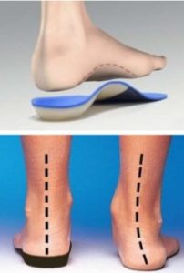
- Arch supports: If predisposing factors involve position abnormalities in the lower limbs or feet, arch supports can have a positive effect on pain.
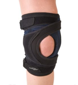
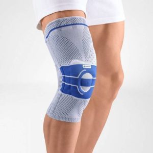
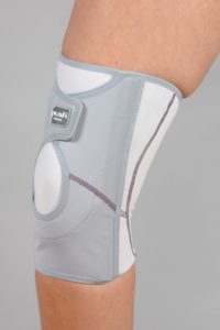
- Bracing: Many patients experience favorable results in pain after wearing a knee brace. However, there is no scientific support that this treatment is strictly necessary. However, exercise therapy with physiotherapy remains the treatment of choice!
- Keyhole surgery of the knee (arthroscopy): Exceptionally, there is a reason to perform keyhole surgery on the knee, for example in case of deterioration after trauma, blockages, frequent effusions... However, due to good imaging techniques, it is no longer necessary to perform keyhole surgery on the knee simply to make a diagnosis.
CARTILAGE INJURIES AND ARTHROSIS OF THE KNEE DISC
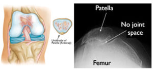
Cartilage damage to the kneecap or the groove in which the kneecap tracks (trochlea) mainly manifests itself through the complaints of patellofemoral pain syndrome. With large cartilage injuries, fluid effusions sometimes occur in the knee, especially after exertion.
Most treatments for patellofemoral pain syndrome are also used for cartilage injuries of the kneecap or the groove of the kneecap. If the examination in the context of the patellofemoral pain syndrome shows that there are cartilage lesions, a number of other treatments are also possible:
Knee injections
The knee is injected with cortisone or hyaluronic acid (gel).
Cortisone (e.g. Depomedrol) is indicated for an acute deterioration with fluid in the knee (post-traumatic, overloaded or inflamed knee).
Hyaluronic acid (gel) (e.g. Ostenil, Synvisc, etc.) can have a positive effect on (morning) stiffness, gnawing and nagging pain in the knee. The injections are usually given in series of three injections shortly after each other to obtain a stronger effect. Active link to hyaluronic acid in general
PRP therapy kan tevens aangeraden worden als bovenstaande inspuitingen in combinatie met andere maatregelen geen effect hebben. Actieve links naar PRP bij algemeen
Keyhole surgery of the knee (arthroscopy)
The cartilage can be stabilized via keyhole surgery with a radiofrequency probe or an ice picking procedure. This works best in case of acute deterioration (post-traumatic) or in case of frequent fluid effusions. Chronic pain complaints respond less well to surgical stabilization.
Patellofemoral prosthesis
If the above treatments do not provide sufficient improvement in the complaints and the cartilage damage is so extensive that the bone of the kneecap largely comes into contact with the bone of the groove, then we speak of terminal osteoarthritis. If there is too much functional hindrance in daily life, a patellofemoral prosthesis may be indicated. The back of the knee and the groove in which the kneecap tracks are then replaced with artificial pieces. The rest of the knee is preserved.
Frequently asked questions

Orthopedisch chirurg – AZ Jan Palfijn Gent
Specialized in shoulder and knee surgery
The information on this page was prepared by Dr. Yves Depaepe and is intended as general medical information. Always consult your doctor for a personalized treatment plan.