DR. YVES DEPAEPE
AC ARTHROSIS or ACROMIO-CLAVICULAR ARTHROSIS
1. WHAT
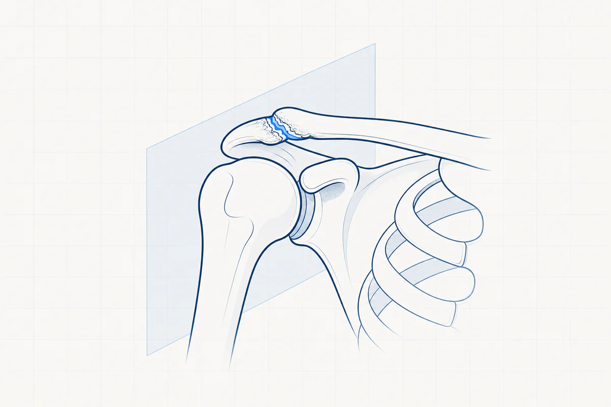
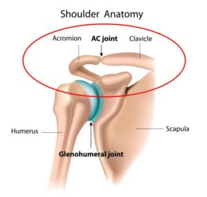
The AC joint is the joint between the acromion (shoulder roof, part of the shoulder blade) and the collarbone (clavicle). The joint ends are covered with cartilage and between them there is a kind of meniscus (cartilage disc).
Both bony prominences are strongly connected to each other by a strong capsule (acromio-clavicular ligaments).
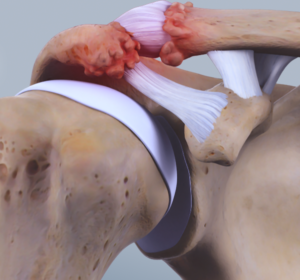
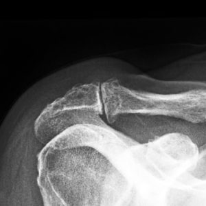
As we age, the joint can become worn. The cartilage wears away, the bone ends rub against each other and ultimately cause pain. This is often accompanied by the development of parrot beaks (osteophytes) on the underside of the joint. These parrot beaks will push on the underlying shoulder tendon and thus cause a kind of clamping.
2. CAUSE
Usually due to progressive wear and tear of the joint.
With long-term physical strain (e.g. years of heavy physical work above shoulder level or years of power lifting), the wear process can be greatly accelerated or a kind of decalcification of the end of the collarbone can occur (osteolysis).
Osteoarthritis can also develop some time after an accident or a fall on the shoulder (post-traumatic osteoarthritis).
3. COMPLAINTS
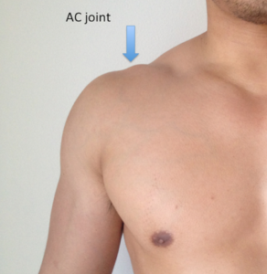
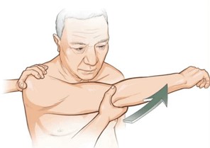
Pain on top of the shoulder, this place can usually be indicated quite correctly. The pain can radiate to the neck or upper arm. The pain usually becomes more intense by lifting loads above shoulder height or by moving the painful arm to the opposite shoulder. Lying on the affected shoulder is often very painful.
Swelling on top of the shoulder due to inflammation or parrot's beak.
Clicking sound on top of the shoulder when moving the arm.
4. EXAMINATIONS AND DIAGNOSIS
Op basis van het verhaal is er vaak al een sterk vermoeden van de diagnose. Bij klinisch onderzoek van de arts is het gewrichtje meestal sterk drukgevoelig. Ook specifieke testen zijn pijnlijk. Klassieke radiografie geeft een idee van de graad van slijtage en de grootte van de papegaaienbekken. Arthro-Ct scan gebeurt soms om ook andere bronnen van schouder pijn op te sporen. Soms is een botscan nodig om de correcte diagnose te kunnen stellen (bv. indien er ook nek afwijkingen aanwezig zijn).
5. TREATMENT
In case of incipient complaints, non-operative treatment is always tried.
NON-OPERATIVE TREATMENT
Rest, avoid actions that provoke pain and avoid heavy loads: do not lift heavy loads above the head.
Anti-inflammatory medication.
Physiotherapy with a focus on restoring mobility, eliminating the inflammation process, learning stretching exercises and learning muscle building exercises.
Injection. A cortisone preparation is usually chosen to control the inflammatory process. This can be repeated two times.
Non-operative treatment is sufficient for a large proportion of patients. In case of persistent, unresolvable complaints or if complaints quickly return, keyhole surgery should be considered.
OPERATIVE TREATMENT = AC resection, mumford operation
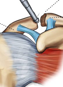
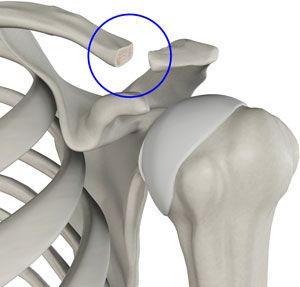
Keyhole surgery is used to perform a complete inspection of the shoulder joint to rule out other causes of shoulder pain. The subacromial space is always enlarged (= subacromial decompression) and all parrot beaks are removed. Afterwards, a piece of the affected, broken cartilage at the end of the collarbone is removed. The space that arises at the end of the collarbone spontaneously fills up with scar tissue.
ANESTHESIA AND HOSPITAL STAY
The keyhole surgery is performed under a short general anesthesia in combination with a local anesthetic (interscalene block).
After the procedure you will stay in the hospital for 1 night.
6. COMPLICATIONS
The most frequent complication is the development of a frozen shoulder (capsulitis or capsule inflammation). This is addressed preventively by quickly starting exercise therapy.
Performing a resection that is too large may cause instability of the collarbone.
Infection, thrombophlebitis or damage to blood vessels or nerves are theoretical complications but in reality they are virtually non-existent.
7. REHABILITATION
To prevent a frozen shoulder, exercise therapy and/or physiotherapy should be started quickly.
The first exercises are taught in the hospital.
A schedule is also provided to start physiotherapy.
During the first 3 weeks, a supporting bandage should be used to control pain and swelling. Afterwards we aim to use the arm without a bandage as quickly as possible within the pain limit. There should be absolutely no heavy strain during the first 6 weeks. We do try to limit all exertion during the first 3 months.
8. FACTS
Try the pain controlled with prescribed painkillers and ice applications.
Love the wounds clean and dry. Showering is permitted with a shower plaster (opsite, tegaderm, etc.). The stitches can be removed after 14 days by the home nurse or your GP.
Disability is between 3 weeks and 4 months and depends on the content of the job. Discuss this with your doctor before the procedure.
Driving. You can start driving when you feel that you can easily steer with both hands without any problem. To do this, you should be able to comfortably raise your arm above shoulder level. For most people this is about three to four weeks after the procedure.
Sports resumption. Walking and cycling are allowed from 3 weeks when the initial pain and swelling have disappeared. Resumption of other sports is individually different and highly sport-specific. This can be discussed with your doctor.
Frequently asked questions

Orthopedisch chirurg – AZ Jan Palfijn Gent
Specialized in shoulder and knee surgery
The information on this page was prepared by Dr. Yves Depaepe and is intended as general medical information. Always consult your doctor for a personalized treatment plan.