DR. YVES DEPAEPE
1. WHAT
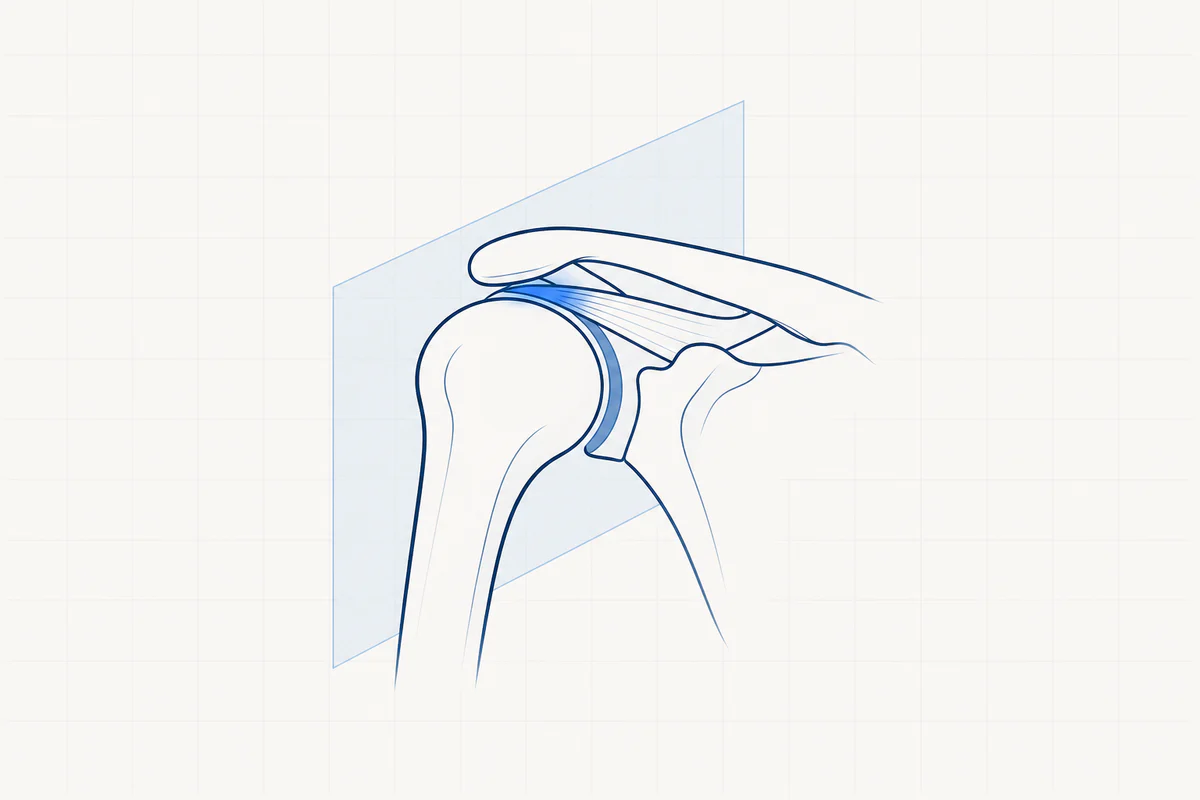
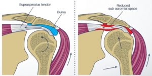
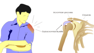
A narrowing of the space under the roof of the shoulder causes the tendons to become clamped between the roof of the shoulder and the head of the upper arm.
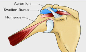
When moving the arm upwards, the tendons of the rotator cuff with the overlying bursa (active link to anatomy) have to slide into a space that is too narrow.
The rubbing of the rotator cuff with the overlying bursa against the roof of the shoulder usually first causes inflammation of the bursa. Continued chafing causes inflammation of the tendon (tendinitis) and persistent irritation can lead to a tear of the rotator cuff tendon.
2. CAUSE
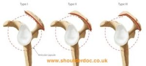
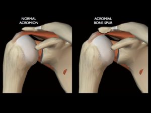
The space between the roof of the shoulder and the upper arm (the subacromial space) may be congenitally narrower. The acromion differs in different individuals (morphological variants). We distinguish 3 types. Type 1 is flat, type 2 is curved and type 3 is angled. Someone with a type 2 or 3 acromion has a higher risk of impingement due to narrowing of the space between the acromion and the humeral head.
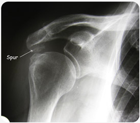
The space can also become narrower over time due to a wear and tear process with bone growth on the underside of the shoulder roof (acromion).
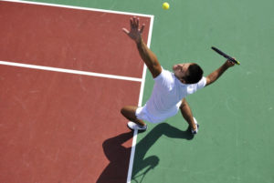
In people who work above shoulder level (e.g. painter) or participate in certain sports (e.g. tennis, volleyball, swimming), this compression will give rise to symptoms more quickly if the subacromial space is already narrower.
3. COMPLAINTS
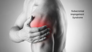
- Pain on the side and front of the shoulder, sometimes the entire upper arm.
- Increased pain when moving the arm upwards, also bringing the arm to the back can be painful.
- Pain during sports movements above the head, such as tennis.
- Pain at night, difficulty sleeping on the shoulder.
- Bij langdurige bestaande pijnklachten kan er krachtsverlies optreden.
4. EXAMINATIONS AND DIAGNOSIS
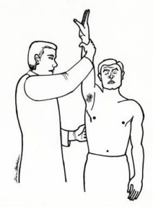
- A good one clinical research by the doctor is essential to suspect the diagnosis.
- One classical radiography gives us a first impression of the distance between the roof and the shoulder ball (subacromial space).
- One ultrasound can demonstrate inflammation of the bursa and/or rotator cuff tendons. Sometimes the arm can be moved during the examination to confirm impingement.
- One MRI scan with contrast (arthro-MRI) can be very useful for further elaboration of the diagnosis.
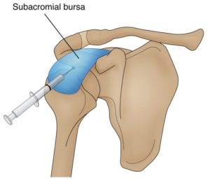
- Sometimes a injection given into the bursa to see if the pain disappears (diagnostic infiltration).
5. TREATMENT
In case of incipient complaints, non-operative treatment is always tried.
NON-OPERATIVE TREATMENT
- Rest, avoiding the actions that provoke pain.
- Anti-inflammatory medication.
- Physiotherapy with a focus on restoring mobility, eliminating the inflammation process, learning stretching exercises and learning muscle-building exercises.
- Injection. A cortisone preparation is usually chosen to control the inflammatory process. This can be repeated two times.
Non-operative treatment is sufficient for a large proportion of patients. In case of persistent, unresolvable complaints or if complaints quickly return, keyhole surgery should be considered.
OPERATIVE TREATMENT = subacromial decompression or acromioplasty
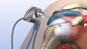
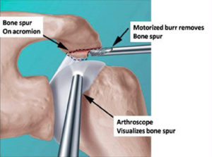
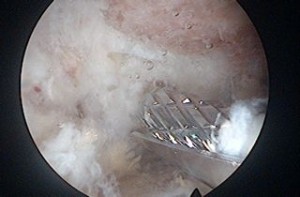
Through keyhole surgery (3 small incisions of 5 mm), the inflamed bursa is removed and the bony spur (bone growth on the roof of the shoulder) is milled away ("shaved"), so that the inflamed tendon has more space and can heal.
During the healing process, a new healthy bursa is formed afterwards.
ANESTHESIA AND HOSPITAL STAY
The keyhole surgery is performed under a short general anesthesia in combination with a local anesthetic (interscalene block).
After the procedure you will stay in the hospital for 1 night.
6. COMPLICATIONS
There are few specific complications with this procedure.
- The most frequent complication is the development of a frozen shoulder (capsulitis or capsule inflammation). This is addressed preventively by quickly starting exercise therapy.
- Infection, thrombophlebitis or damage to blood vessels or nerves are theoretical complications but in reality they are virtually non-existent.
7. REHABILITATION
To prevent a frozen shoulder, exercise therapy and/or physiotherapy should be started quickly.
The first exercises are taught in the hospital.
A schedule is also provided to start physiotherapy.
A carrying bandage can only be used for pain during the first 3 weeks. It is not strictly necessary to wear this full time.
We aim to use the arm without a bandage as quickly as possible within the pain limit.
We do try to limit all exertion during the first 3 months.
8. FACTS
- Try the pain controlled with prescribed painkillers and ice applications.
- Love the wounds clean and dry. Showering is permitted with a shower plaster (opsite, tegaderm, ..) The stitches can be removed after 14 days by the home nurse or your GP
- Disability is between 2 weeks and 4 months and depends on the content of the job. Discuss this with your doctor before the procedure.
- Driving. You can start driving when you feel that you can easily steer with both hands without any problem. To do this, you should be able to comfortably raise your arm above shoulder level. For most people this is about three to four weeks after the procedure.
- Sports resumption. Walking and cycling are allowed from 3 weeks when the initial pain and swelling have disappeared. Resumption of other sports is individually different and highly sport-specific. This can be discussed with your doctor.
Frequently asked questions
Orthopedisch chirurg – AZ Jan Palfijn Gent
Specialized in shoulder and knee surgery
The information on this page was prepared by Dr. Yves Depaepe and is intended as general medical information. Always consult your doctor for a personalized treatment plan.