DR. YVES DEPAEPE
AC LUXATION or ACROMIO-CLAVICULAR LUXATION
1. WHAT
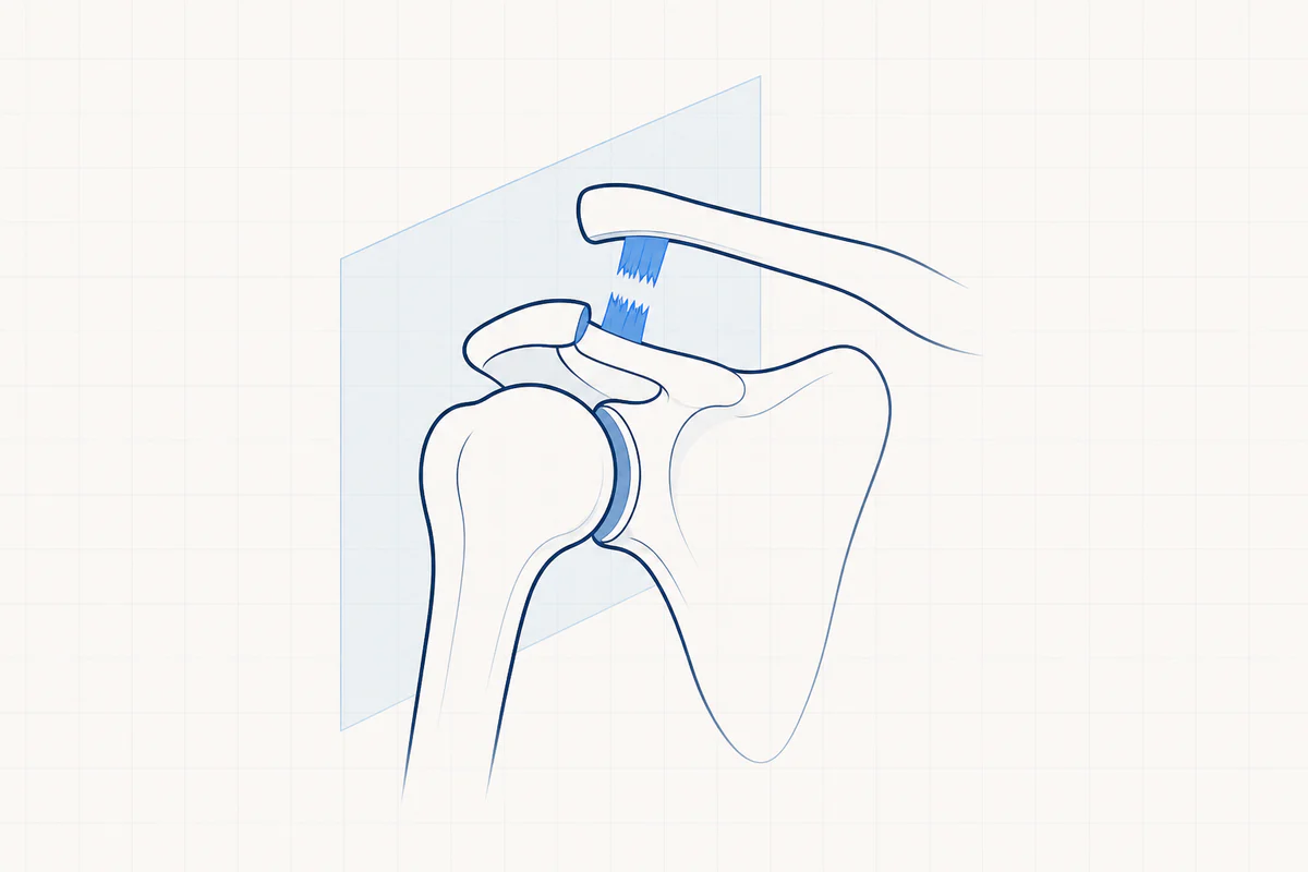
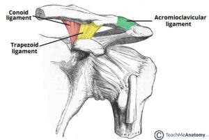
The AC joint is the joint between the acromion (shoulder roof, part of the shoulder blade) and the collarbone (clavicle). The joint ends are covered with cartilage and between them there is a kind of meniscus (cartilage disc).
Both bony prominences are strongly connected to each other by a strong capsule (acromio-clavicular ligaments). The collarbone is additionally stabilized by 2 ligaments in the front between the coracoid process (anterior protrusion of the shoulder blade) and the collarbone (trapezoid ligament and coronoid ligament).
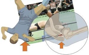
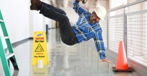
A fall on the shoulder or a fall on an outstretched arm can cause a dislocation of the AC joint. Because the ligaments tear, the collarbone can move upwards and a stair position forms at the end of the collarbone.
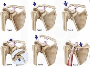
Six types of luxations are distinguished, according to the severity of the injuries
- Grade 1: Contusion/strain of the AC joint without true ligament tears.
- Grade 2: Tear of the capsule surrounding the AC joint.
- Grade 3: Also tear of two other ligaments (coronoid and deltoid ligament)
- Grade 4: The collarbone also perforates the muscles, which are partly torn.
- Grade 5: The same, with large displacement and significant tearing of the muscles
- Grade 6: The same, but with displacement of the collarbone downwards.
2. CAUSE
Trauma, fall.
3. COMPLAINTS
- Pain varies depending on the severity of the injury
- Swelling on top of the shoulder
- Blue to yellowish discoloration at the front of the shoulder
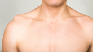
- Visible kick position on top of the shoulder
4. EXAMINATIONS AND DIAGNOSIS
Bij klinisch onderzoek is de trapstand duidelijk merkbaar. Bij druk op het uiteinde van het sleutelbeen kan dit naar beneden geduwd worden, wanneer we het sleutelbeen weer loslaten, zien we het uiteinde onmiddellijk terug naar boven schieten (piano toets teken). Radiografie toont ons de graad van de ontwrichting en/of eventuele breuken. In zeldzame gevallen is er bijkomend noodzaak tot echografie of CT scan.
5. TREATMENT
All grade 1 and grade 2 injuries and some grade 3 injuries are treated non-operatively.
NON-OPERATIVE TREATMENT
- Shoulder bandage or sling for 3 weeks.
- Anti-inflammatory medication and painkillers.
- Physiotherapy focusing on restoring mobility can usually start after about 3 weeks.
- Do not lift or lift during the first 6 weeks.
Some grade 3 injuries and almost all grade 4, 5 and 6 injuries are treated surgically.
OPERATIVE TREATMENT
The collarbone is surgically returned to the correct height and held in place with pins, wires, screws, angle plates, tendon, etc.
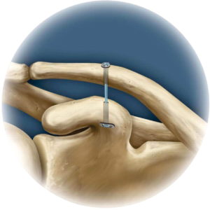
We usually opt for the use of recent disruptions a tight rope. This is a thick cord that is stretched between the collarbone and the coracoid protrusion and is tensioned on two knots. This brings the collarbone back to the correct height and allows the damaged ligaments to recover to the correct length. If the cartilage of the joint is additionally damaged, the end of the collarbone is also removed (AC resection).
In case of late detection and chronic injuries, we also use our own tendon to stabilize the collarbone.
ANESTHESIA AND HOSPITAL STAY
The procedure is performed under a short general anesthesia in combination with a local anesthetic (interscalene block).
After the procedure you will stay in the hospital for 1 night.
6. COMPLICATIONS
- Persistent pain complaints.
- Aesthetically lasting elevation of the collarbone.
- Slight loss of strength.
- Loosening of the fixation of the material or tendon used.
- Infection risk is reduced by administering antibiotics during the procedure.
- Damage to blood vessels or nerves are theoretical complications, but in reality they are virtually non-existent.
7. REHABILITATION
Om een frozen shoulder te voorkomen wordt snel met oefentherapie gestart. De eerste oefeningen (pendelen) worden in het ziekenhuis aangeleerd. Gedurende de eerste 3 weken moet gebruik gemaakt worden van een draagverband om de heling van de ligamenten de beste kans te geven. Gedurende de eerste 6 weken mag er absoluut geen zware belasting zijn en proberen we alle beweeglijkheid boven het niveau van de schouder te vermijden. Er wordt bijkomend een schema meegegeven om kinesitherapie te starten zo nodig vanaf 3 weken.
8. FACTS
- Try the pain controlled with prescribed painkillers and ice applications.
- Love the wounds clean and dry. Showering is permitted with a shower plaster (opsite, tegaderm, ..) The stitches can be removed after 14 days by the home nurse or your GP
- Disability is between 3 weeks and 4 months and depends on the content of the job. Discuss this with your doctor before the procedure.
- Driving. You can start driving when you feel that you can easily steer with both hands without any problem. To do this, you should be able to comfortably raise your arm above shoulder level. For most people this is about four to six weeks after the procedure.
- Sports resumption. Walking and cycling are allowed from 3 weeks when the initial pain and swelling have disappeared. Resumption of other sports is individually different and highly sport-specific. This can be discussed with your doctor.
- It material (tight rope) can remain in place and does not have to not to be removed.
Frequently asked questions
Orthopedisch chirurg – AZ Jan Palfijn Gent
Specialized in shoulder and knee surgery
The information on this page was prepared by Dr. Yves Depaepe and is intended as general medical information. Always consult your doctor for a personalized treatment plan.