DR. YVES DEPAEPE
1. WHAT
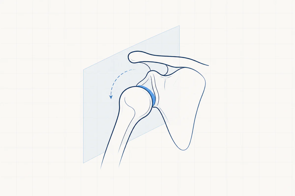
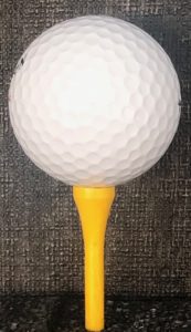
The shoulder joint is a very mobile joint. We can compare the shoulder ball and the shoulder socket to a golf ball on a tee. The price we pay for this great flexibility is "instability".
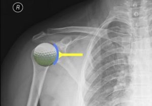
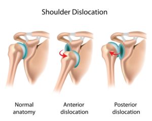
Instability is the inability to keep the bulb (humeral head) centered in the socket (glenoid). If the joint is unstable, this leads to a dislocation (luxation) of the shoulder. Sometimes the shoulder also threatens to dislocate, but this does not lead to complete dislocation (subluxation).
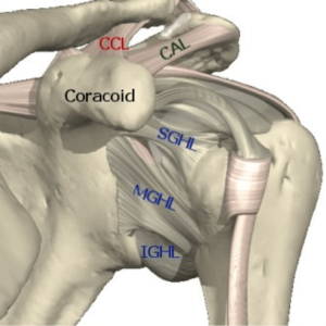
To guarantee the stability of the shoulder in normal circumstances, we have a sturdy membrane around the shoulder (capsule) containing strong reinforcing ligaments (ligaments). In addition, there is a firm upright, cartilaginous ring around the socket, to which the capsule attaches. The rotator cuff muscles (active link to anatomy) also provide additional dynamic stability, with the rotator cuff keeping the ball centered in the socket.
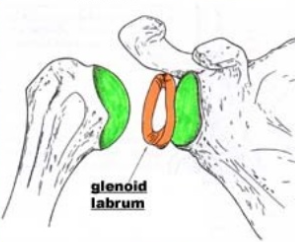
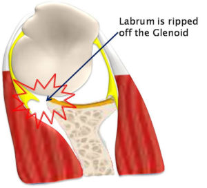
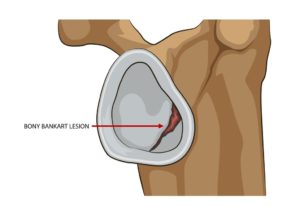
In the majority of cases (95%) the head dislocates anteriorly (anterior luxation), in a limited number of cases (5%) the head dislocates posteriorly (posterior luxation). If the shoulder dislocates, the labrum with the capsule on it can tear away from the socket (Bankart injury). Sometimes it can even break off a piece of bone in the socket (bony bankart injury).
Bijkomend ontstaat er een indeuking in de bol (humeruskop) van de schouder omdat deze tijdens de ontwrichting tegen de rand van de kom (glenoid) wordt geduwd (hill sachs letsel). Bij oudere mensen kan een ontwrichting ook gepaard gaan met een scheur in de rotator cuff.
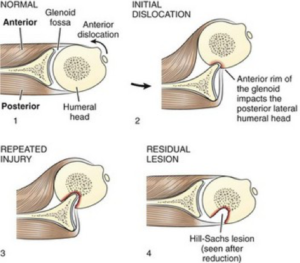
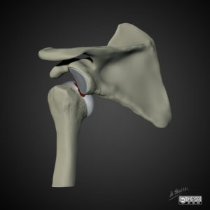
When the shoulder is dislocated, nerves can sometimes be stretched. This recovery can often take months.
2. CAUSE
The traumatic luxation
The most common cause of dislocation is after an accident. When sufficient forces are placed on the shoulder joint due to trauma, the shoulder can be pushed into an extreme position and pulled out of its socket. A fall during a football match could be an example.
The non-traumatic luxation
This occurs when the shoulder dislocates with minimal force such as reaching for an object or turning over in bed. Usually it will pop back in on its own or with a little help. This type of luxation mainly occurs in people with hyperlax joints, for example people who can overextend their knees and elbows. The firm capsule and the underlying ligaments are naturally "looser".
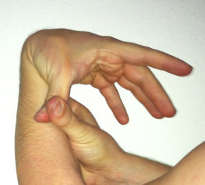
Acquired instability
Through actions or sports in which the shoulder is frequently put in extreme positions, the capsule and ligaments are stretched little by little. This can gradually cause a dislocation.
3. COMPLAINTS
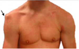
In the event of a dislocation, sudden, excruciating pain occurs and you can no longer move the shoulder. The shape of the shoulder changes and looks "abnormal". With a partial dislocation (subluxation), this usually feels like a vague pain, accompanied by a click.
4. EXAMINATIONS AND DIAGNOSIS
A good clinical examination by the doctor is essential to suspect the diagnosis and to detect whether there may be accompanying nerve injuries. A classic radiography shows the dislocation and also any additional injuries, such as fractures. In the second phase, and especially in the case of recurrent dislocations, a CT scan or MRI scan with contrast will also be planned to visualize the damage to the capsule, ligaments and labrum.
- One good clinically Examination by the doctor is essential to suspect the diagnosis and to detect whether there may be accompanying nerve injuries.
- One classical radiography shows the dislocation and also any additional injuries such as fractures
- In the second phase and especially in the case of recurrent dislocations, an additional CT scan or MRI scan can be planned with contrast to visualize damage to the capsule, ligaments and labrum.
5. TREATMENT
During emergency admission or in the operating room, the shoulder is put back into place (reduction), with or without light anesthesia. In patients with chronic, recurrent instability, they are often able to push the shoulder back into place on their own.
After reduction, a shoulder brace will be applied, usually for about three weeks. During this period, an appointment will be made with your treated shoulder specialist.
Physiotherapy is necessary as a first treatment to regain mobility and to learn muscle strengthening exercises.
Further treatment depends on the diagnosis and any lesions found on imaging. The aim of further treatment is to repair the damage that has occurred, but also to ensure that new dislocation can be avoided.
If there have already been several dislocations, there is a greater chance that damage has already occurred in the shoulder joint (e.g. Bankart injury). The younger you were at the time of the first dislocation, the greater the chance that additional dislocations will follow. The more dislocations there have been, the greater the chance of damage to the joint and the greater the chance of osteoarthritis later in life. Your sporting or professional activities can also make you susceptible to new dislocations and can be decisive for further treatment (e.g. a rugby player has a greater chance of new dislocations than a runner).
NON-OPERATIVE TREATMENT
- Rest, shoulder brace in the first 3 weeks after dislocation.
- Anti-inflammatory medication.
- Physiotherapy with a focus on restoring mobility, eliminating the inflammation process and learning muscle-building exercises.
- In the case of non-traumatic instability and congenital instability, there will be a need for very long-term physiotherapy to obtain sufficient muscle strengthening.
Non-operative treatment is sufficient for a large proportion of patients. In case of persistent, unresolvable complaints or if complaints quickly return, keyhole surgery should be considered.
OPERATIVE TREATMENT
Keyhole surgery: arthroscopic bankart repair
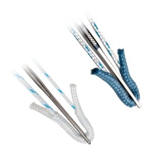
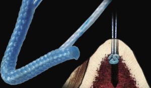
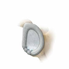
During keyhole surgery, the torn labrum and capsule are reattached to the bone of the socket (glenoid) using special anchors with wire. This suture also tightens the ligaments. These anchors no longer need to be removed.
If the "indentation" in the head of the upper arm (Hill Sachs injury) is too large, an anchor is placed in the dent as additional reinforcement and the capsule and part of the rotator cuff are sutured into it, a so-called remplissage.
Latarjet procedure
Deze open ingreep (met insnede van 8 tal centimeter vooraan op de schouder) wordt meestal uitgevoerd als er bijkomend een stukje bot is afgebroken van de kom of als de schouder opnieuw ontwricht na een vroeger uitgevoerde kijkoperatie. In sommige gevallen wordt dit als eerste keuze operatie uitgevoerd bv. atleten in contactsporten zoals rugby, mensen met een zwaar schouder belastend beroep.
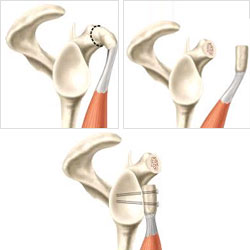
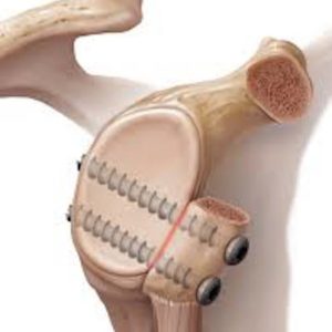
The front bony protrusion of the shoulder blade (coracoid process) is detached and, together with the tendons on it, transplanted to the front of the socket of the shoulder (glenoid). This piece of bone is secured with 2 screws. The piece of bone provides a larger surface area for the socket, the tendons attached to it provide additional strength so that the shoulder can no longer come out of the socket.
If the screws cause discomfort after the piece of bone has grown together, they can be removed.
Bone block procedure
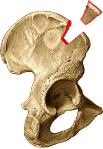
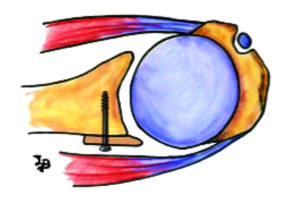
After recurrent posterior dislocations after suturing via keyhole surgery, bone loss after a posterior dislocation or recurrent dislocation after a latarjet procedure, a bone block is placed against the socket of the shoulder. A piece of bone measuring 3x2cm is removed at the level of the iliac crest and then screwed into place at the level of the injury to the socket. This way the surface of the bowl is made larger.
ANESTHESIA AND HOSPITAL STAY
The keyhole surgery is performed under a short general anesthesia in combination with a local anesthetic (interscalene block). After the procedure you will stay in the hospital for 1 night.
6. COMPLICATIONS
There is no such thing as a procedure without possible complications, but overall the complications are very limited.
However, the chance of lasting stability after a correctly performed operation is never 100%!
- Disrupt again. With suturing via keyhole surgery, the risk is around 15%. The risk is greatest, especially in young patients involved in competitive contact sports. With an open latarjet procedure, the chance of recurrence is around 5%.
- Infection is almost non-existent during keyhole surgery, with an open latarjet procedure there is a very limited risk. This is anticipated by administering an antibiotic during the procedure.
- Decreased mobility. In the initial phase there is always a reduced ability to rotate the arm outward (external rotation). This disappears spontaneously with time.
- Thrombophlebitis or damage to blood vessel or nerve. are theoretical complications but in reality very rare.
7. REHABILITATION
Na de operatie draagt u gedurende 4 tot 6 weken een schouder brace. Het vastgemaakte labrum en/of het bot moet terug vastgroeien aan het bot van de kom van de schouder. Dit proces neemt ongeveer een 3 tal maanden in beslag en gedurende deze periode is de schouder kwetsbaar.
The physiotherapy is done according to a given schedule. The timing of the start of physiotherapy depends on the size of the injury and the surgical technique performed. We often wait 2 to 4 weeks before starting the physiotherapy treatment. During your hospital stay, you will be taught exercises that you can perform independently at home.
We do try to limit all exertion during the first 3 months.
Overall, the speed of rehabilitation from a Latarjet procedure is slightly faster than an arthroscopic Bankart recovery.
8. FACTS
- Try the pain controlled with prescribed painkillers and ice applications.
- Love the wounds clean and dry. Showering is permitted with a shower plaster (opsite, tegaderm, ..) The stitches can be removed after 14 days by the home nurse or your GP.
- Disability. is between 6 weeks and 4 months and depends on the content of the job. Discuss this with your doctor before the procedure.
- Driving. You can start driving when you feel that you can easily steer with both hands without any problem. To do this, you should be able to comfortably raise your arm above shoulder level. For most people this is about 6 to 8 weeks after the procedure.
- Sports resumption. Your physiotherapist and surgeon will advise you when it is safe to resume your hobbies. This will depend on the type of sport and the level at which you practice it.
- Weeks 1 to 6: no sports, only entertaining cardio training can be allowed (e.g. exercise bike or treadmill).
- Week 6 to 16: walking, cycling and swimming (breaststroke first) can be gradually built up within the pain threshold.
- Week 16 to 24: sport-specific training for contact sports and ball sports can be started.
- From 6 months After the procedure, a return to competition can be planned.
Frequently asked questions
Orthopedisch chirurg – AZ Jan Palfijn Gent
Specialized in shoulder and knee surgery
The information on this page was prepared by Dr. Yves Depaepe and is intended as general medical information. Always consult your doctor for a personalized treatment plan.