DR. DEPAEPE
1. WHAT
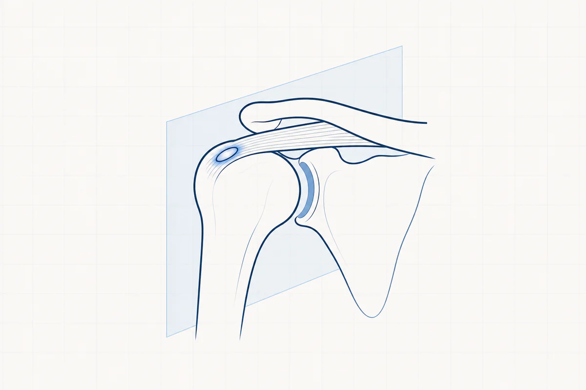
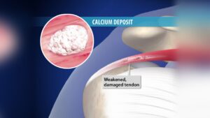
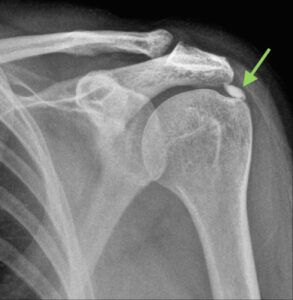
It concerns a deposit of calcium crystals in one or more tendons of the rotator cuff. This calcium deposit is rather soft (like toothpaste) and looks white on radiography. The rotator cuff is the most affected tendon where calcium deposits. Not everyone with calcium deposits in the shoulder is aware of this or experiences complaints.
In some cases the body has the opportunity to dissolve the lime deposit (dissolution phase), in other cases the inflammatory process continues without dissolving the lime.
2. CAUSE
The cause is very poorly known. Calcifications mainly occur in areas of the tendon with reduced blood supply. Genetic factors also play a role. Calcification is not the result of an accident or dietary habits, arthrosis or osteoporosis.
3. COMPLAINTS
Complaints vary depending on the stage of the disease.
There are few complaints in the phase when the calcification is growing. If the volume increases, a classic clamping or impingement problem arises. The volume narrows the space between the roof of the shoulder and the tendon of the shoulder, causing friction of the tendon against the roof, resulting in irritation and inflammation.
The pain is mainly located on the side of the upper arm. The pain will increase with activities above shoulder level (e.g. getting something from a high cupboard) and when bringing the hand to the back (internal rotation). There is also very frequent night pain when lying on the shoulder.
As mentioned, the body sometimes tries to dissolve the calcification (= solution phase). The calcium will be released in the bursa (between the rotator cuff and the shoulder roof) and cause very acute pain with very severe limitation of mobility.
4. EXAMINATIONS AND DIAGNOSIS
- The suspicion of calcification is retained during the examination by your doctor.
- An X-ray clearly shows the large calcifications.
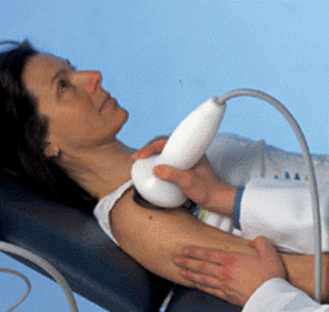
- Ultrasound clearly shows the large and smaller (microcalcifications) calcifications.
- An arthro-CT shows the calcification in a 3D volume and also provides more information about the integrity of the tendon.
- Arthro-MRI is a good examination to rule out other causes of the pain, but it does not visualize the calcification itself as well.
5. TREATMENT
The course of calcification is very unpredictable. Sometimes we also see a spontaneous disappearance of the calcium deposit, usually accompanied by a severe pain flare (dissolution phase).
NON-OPERATIVE TREATMENT
- Treat the symptoms by relative rest, taking painkillers and anti-inflammatories.
- If this is insufficient, 2 cortisone infiltrations can be administered, especially with the aim of eliminating the inflammation. These infiltrations are certainly useful in the acute, painful phase (resolution phase). Limiting to 2 infiltrations will ensure that there are no adverse effects on bone or tendons.
- Supportive physiotherapy can also be followed, mainly anti-inflammatory physiotherapy and physiotherapy to maintain mobility.
- Needling: mainly performed by radiologists, less popular in Belgium. The calcium depot is punctured under local anesthesia under radioscopic control. It is hoped to remove some of the limescale and in addition, increased blood flow is created around the calcification.
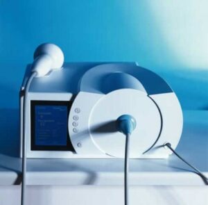
- ESWT: extracorporeal shock wave therapy. This technique was developed by analogy with the kidney stone crusher. The technique uses focused sound waves. However, the calcification is not “smashed” here (after all, it is a soft structure, like toothpaste). In particular, the blood supply in the surrounding tissues is stimulated so that a better anti-inflammatory effect is achieved and the possible resolution phase can be accelerated. This treatment is not painless and is best done under local anesthesia. In practice, we notice that a maximum of 50% of patients benefit from ESWT treatment. ESWT is not recommended shortly after injection with corticoids, with osteosynthesis material present, with a pacemaker, with clotting problems, etc.
OPERATIVE TREATMENT – ARTHROSCOPY
If previous therapy fails or if there is too much pain, keyhole surgery should be performed.
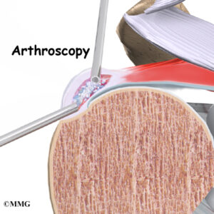
The most common way to remove calcium deposits is via arthroscopy (keyhole surgery). The procedure is performed under a short general anesthesia, often in combination with local anesthesia (interscalene block).
The calcium is detected with the camera, punctured with a needle and then the tendon is incised superficially so that the calcium can be flushed out (such as pushing out an abscess/ulcer) and the tendon can be scraped out (curettage). Afterwards, the inflamed bursa is removed and a “decompression” is performed: a few millimeters of bone are removed with a burr from the underside of the acromion (roof of the shoulder) to give the swollen tendon more space.
The major advantage of performing the procedure via keyhole surgery is that if other injuries are detected at the same time, they can be treated in the same procedure.
6. COMPLICATIONS AFTER PROCEDURE
There are few specific complications with this procedure.
The most frequent complication is the development of a frozen shoulder (capsulitis).
This is addressed preventively by starting rapid exercise therapy.
7. REHABILITATION AFTER OPERATION
To prevent a frozen shoulder, exercise therapy and/or physiotherapy should be started quickly.
The first exercises are taught in the hospital.
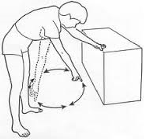
A schedule is also provided to start physiotherapy.
A carrying bandage can only be used for pain during the first 3 weeks.
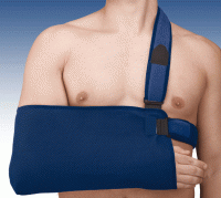
We aim to use the arm without a bandage as quickly as possible within the pain limit.
The total recovery time is between 6 and 12 weeks and mainly depends on the size of the lime deposit. After all, the remaining cavity must close with a scar.
The disability will depend on the profession and is between 1 and 4 months.
8. FACTS
- Try the pain controlled with prescribed painkillers and ice applications.
- Love the wounds clean and dry. Showering is permitted with a shower plaster (opsite, tegaderm, ..). The stitches can be removed after 14 days by the home nurse or your GP.
- Disability is between 2 weeks and 3 months and depends on the content of the job. Discuss this with your doctor before the procedure.
- Driving. You can start driving when you feel that you can easily steer with both hands without any problem. To do this, you should be able to comfortably raise your arm above shoulder level. For most people this is about four weeks after the procedure.
- Sports resumption. Walking and cycling are allowed from 3 weeks when the initial pain and swelling have disappeared. Resumption of other sports is individually different and highly sport-specific. This can be discussed with your doctor.
Frequently asked questions
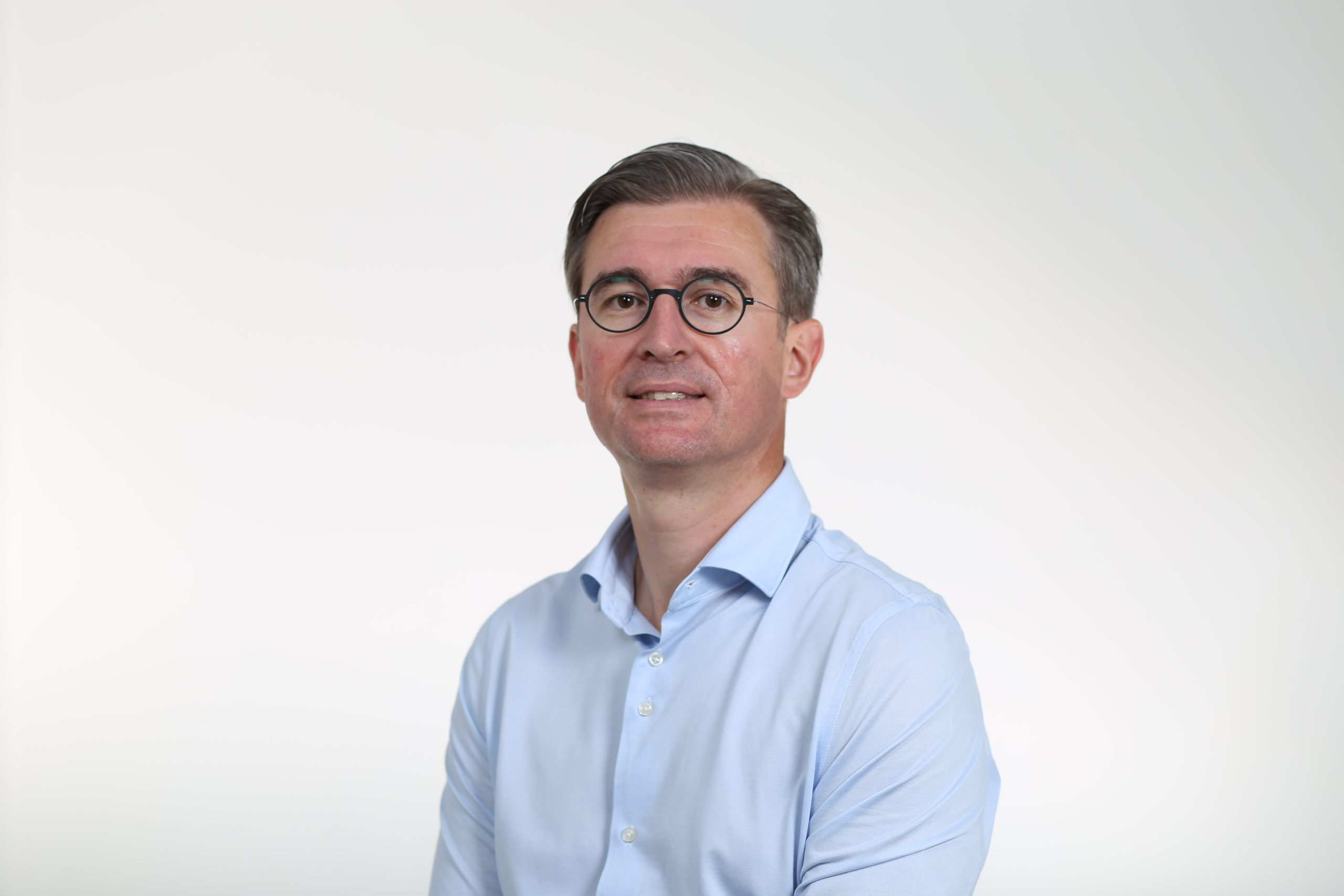
Orthopedisch chirurg – AZ Jan Palfijn Gent
Specialized in shoulder and knee surgery
The information on this page was prepared by Dr. Yves Depaepe and is intended as general medical information. Always consult your doctor for a personalized treatment plan.