DR. YVES DEPAEPE
A. Shoulder inflammation or arthritis of the shoulder joint
There are 5 different causes for the development of arthritis of the shoulder. These conditions may require the placement of a shoulder prosthesis if pain persists and increases in functional impairment.
1. Arthrosis of the shoulder
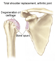
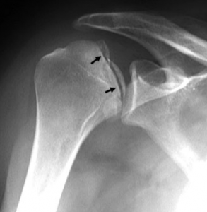
Osteoarthritis of the shoulder is a chronic wear and tear process in which the protective cartilage at the bone ends gradually decreases in volume and quality. In addition to the reduction of cartilage, the quality of the tissue of the shoulder tendons also decreases with age, making them less able to perform their movement function.
As a result of the damage to the cartilage, the bony ends rub directly against each other, causing pain. In addition, swelling, stiffness and deformities occur.
This leads to loss of strength and often causes functional limitations in daily activities (e.g. getting something out of the cupboard, pouring coffee, washing, etc.).
2. Rheumatoid arthritis
In patients with inflammatory rheumatism or rheumatoid arthritis (RA), accelerated cartilage breakdown and frequent inflammation in the joints occur. There is usually a typical symmetrical involvement of the joints.
Rheumatoid arthritis is an autoimmune disease. The cause of this condition is a fault in the immune system.
As the cartilage breaks down, this leads to pain in the shoulder and significant limitation of function.
3. Trauma
After a fracture or dislocation of the shoulder, accelerated arthrosis of the shoulder joint can develop after a certain time.
4. Avascular necrosis
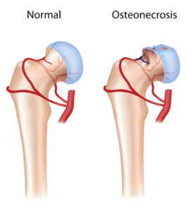
This is a disease that occurs when the blood supply to the head of the humerus is interrupted. The area of bone in the humeral head that does not receive blood will die (necrosis). This ultimately leads to deformation of the shoulder joint and the development of osteoarthritis.
Some causes for the development of avascular necrosis are long-term cortisone use, excessive alcohol use, fractures, ... Sometimes no cause is identified: idiopathic avascular necrosis.
5. Rotator cuff arthropathy
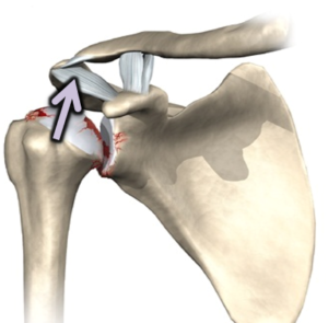
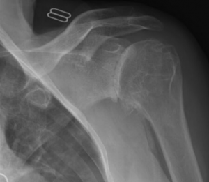
If a rotator cuff tear has not been repaired for a long time, significant osteoarthritis can develop. The rotator cuff can no longer center the ball of the shoulder in the pan of the shoulder. The ball of the shoulder can rise and rub against the roof of the shoulder. This causes secondary arthrosis of the shoulder.
The combination of a rotator cuff tear and significant osteoarthritis of the shoulder leads to pain and sometimes very significant limitation of movement and muscle weakness. This is called a "pseudoparalysis" image: it appears as if the arm is paralyzed.

B. Fractures of the shoulder
Fractures of the ball of the shoulder (humeral head) are common.
In the majority of cases, these fractures will not require surgery.
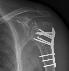
Sometimes these fractures need to be treated with an operation in which the pieces are put back in the right place (osteosynthesis with plate or nail).
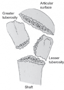
However, sometimes it is impossible to piece the various fracture fragments back into the right place and fix them together with screws, and then it is appropriate to place a shoulder prosthesis.
Diagnosis
A clinical examination will be performed during the consultation. Further imaging will be selected based on this research.
In addition to standard radiographic images, an (arthro) CT scan and/or an (arthro) MRI scan of the shoulder may also be performed.
Non-surgical treatments
- Physiotherapy can maintain the mobility of the shoulder.
- Pain medication and anti-inflammatory medications. This medication is often stomach-irritating and cannot be taken for a long time.
- Injections:
- Corticosteroids: to control the acute inflammatory process. Quick and only temporary effect.
- Hyaluronic acid infiltrations
- PRP infiltrations
- Dietary supplements e.g. glucosamines and chondroitin. There is very little scientific support for the effectiveness of these products in shoulder osteoarthritis.
When which shoulder prosthesis? Types of prosthetics
Which shoulder prosthesis is most suitable for you depends on the underlying diagnosis. In consultation with you, your doctor will choose the prosthesis that is most suitable for your situation.
1. Reverse shoulder prosthesis
This is the most frequently performed shoulder prosthesis.
If not only the shoulder joint, but also the tendons of the shoulder are worn out, a reverse shoulder prosthesis will be fitted.
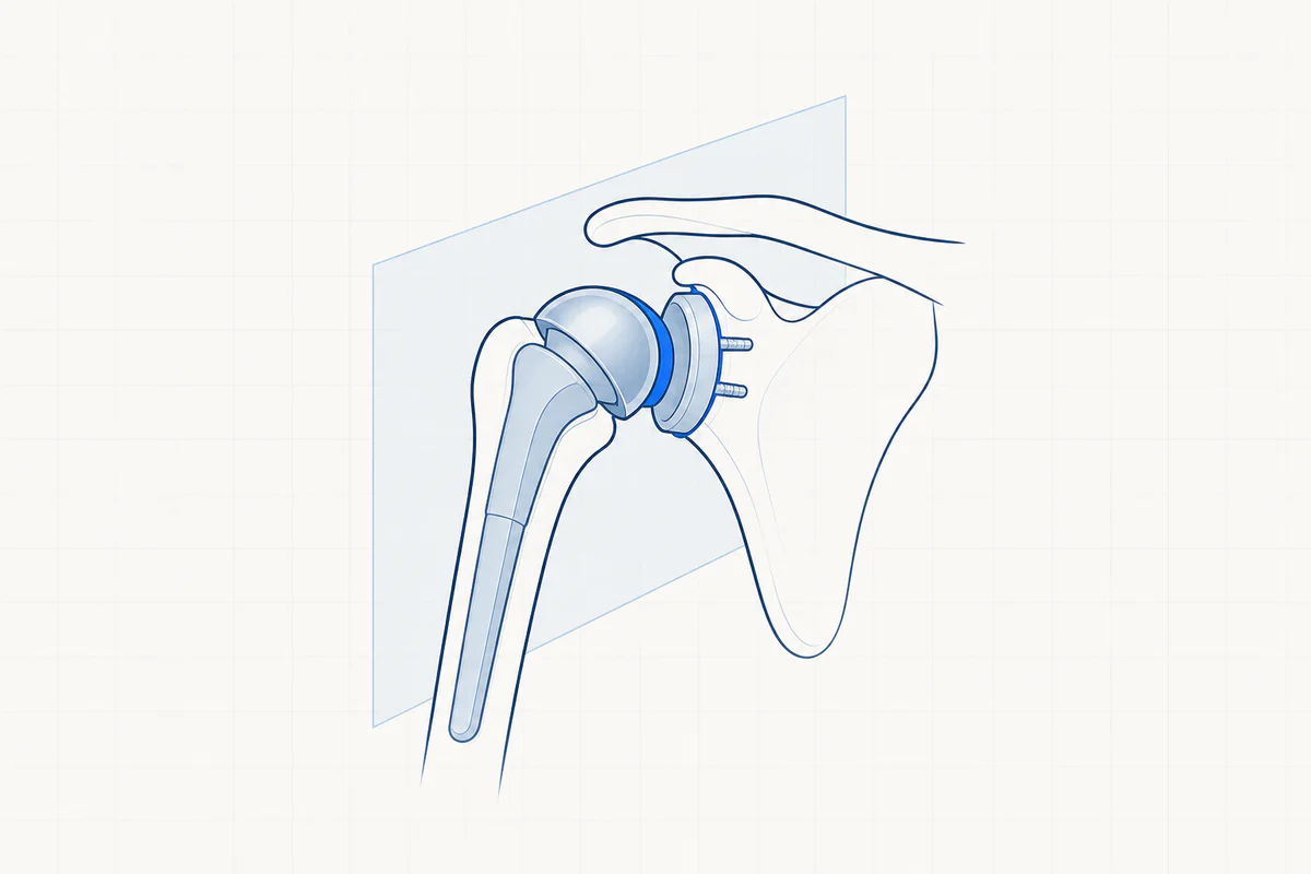
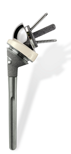
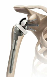
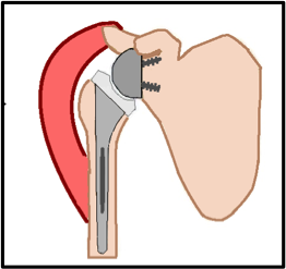
With a reverse shoulder prosthesis, a hemisphere is placed in the place of the original pan. The original ball of the shoulder is then replaced by a socket that is attached to the stem. The parts of the prosthesis are therefore placed "inverted", as it were.
This prosthesis completely changes the biomechanics of the shoulder. The rotator cuff tendons for lifting your arm become very important. The motor of the prosthesis is the large shoulder muscle (deltoid muscle). The deltoid muscle together with the reverse shoulder prosthesis ensure that you can raise the arm again, even with a torn rotator cuff tendon.
2. Anatomical shoulder prosthesis
With an anatomical shoulder prosthesis, the worn ball of the shoulder (the humeral head) is sawed off and replaced with a new ball. This is placed on a stem, such that the head can grow firmly into the bone to guarantee optimal stability. The shoulder pan (the glenoid) is replaced by polyethylene (a hard plastic).
An anatomical shoulder prosthesis is usually placed in younger patients with osteoarthritis. An anatomical shoulder prosthesis can only be placed if all structures of the rotator cuff tendons are still completely intact.
Note: voiceless shoulder prosthesis
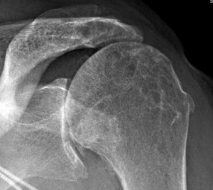
With a stemless shoulder prosthesis, a metal ball is placed with a much shorter stem or even no stem. A stemless shoulder prosthesis can only be placed if the bone quality is optimal.
3. Fracture of prosthesis
This type is used for irreparable fractures. In the vast majority of cases this concerns a reverse shoulder prosthesis. It is rarely opted to replace only the broken ball: half shoulder prosthesis (hemiprosthesis).
The procedure is more complex than a prosthesis for the treatment of osteoarthritis and the result is more difficult to predict. In many cases we cannot achieve a complete but acceptable recovery of shoulder function. The rehabilitation is more difficult and often longer than with a classic reverse shoulder prosthesis.
Complications
Possible risks and complications that may occur with this procedure:
- An infection or contamination of the prosthesis
- Internal or external bleeding
- Dislocation of the prosthesis (dislocation)
- Injuries to blood vessels or nerves
- Stiffness after the procedure with a loss of mobility of the shoulder
- A fracture of the shoulder blade due to overload
- A fracture around the prosthesis
- Premature loosening or wearing out of the prosthesis at an early stage
If you have any questions about the complications, discuss this with your treating physician. Please note that a shoulder prosthesis has now become a routine procedure and the chance of complications has become very small (but this chance can never be reduced to 0).
Facts - Aftercare after shoulder prosthesis
- In the first 4 weeks the arm is kept in a sling. Specific instructions will be given to you upon discharge, as the duration may vary depending on the type of prosthesis.
- In the first 3 weeks After the procedure there is a need for wound care. The sutures can be removed after 14 days (usually dissolvable sutures).
- It is useful daily and frequently to lay ice on the shoulder.
- The physiotherapy is started between weeks 1 and 4. The time depends on the type of prosthesis. You will receive a prescription with a schedule from the treating surgeon.
- From day 1, light exercises (commuting exercises) performed. These are learned during a hospital stay.
- The first 6 weeks are mainly aimed at restoring passive mobility. The second period in between 6 and 12 weeks is mainly aimed at restoring active mobility.
- Strength exercises are only important once mobility has been sufficiently recovered.
- The total rehabilitation is very different from person to person and strongly depends on the type of prosthesis: a fracture prosthesis will have much slower rehabilitation than a planned reversed prosthesis.
- The total recovery time is between 6 and 12 months.
Frequently asked questions

Orthopedisch chirurg – AZ Jan Palfijn Gent
Specialized in shoulder and knee surgery
The information on this page was prepared by Dr. Yves Depaepe and is intended as general medical information. Always consult your doctor for a personalized treatment plan.